Helminths can inhabit a person in almost all organs. In accordance with this, the ways of their penetration into the human body, the symptomatology of diseases and diagnostic methods are different.
20.1. TYPE FLAT WORMPLA THELMINTHES
Flatworms have a body that is flattened in the dorsoventral direction. There is no body cavity, internal organs are immersed in loose connective tissue - parenchyma. The musculocutaneous sac consists of integumentary tissue - the tegument, which is a multinucleated non-cellular structure, and three layers of smooth muscles - longitudinal, transverse and dorsoventral. Their movements are slow and imperfect. Nervous system consists of nerve nodes at the anterior end of the body, from which longitudinal nerve cords extend posteriorly. Digestive system, if present, it is built up of the pharynx and intestines, which are blindly closed. Undigested food debris is passed through the mouth. Reproductive system hermaphrodite and very complex built.
Selection is done using protonephridial system, consisting of separate excretory cells - proteonephridia. They are able to capture dissimilation products and transport them along intracellular channels running in their long processes. The products of excretion enter the collecting ducts, and from there either directly or through the bladder into the external environment.
Species of medical importance are presented in two classes: Flukes and Tapeworms.
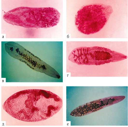
20.1.1. PUCKER CLASS TREMATODA
digestion products. Otherwise, the flukes repeat the organization of the flatworms described above (Figure 20.1).
Miradiations enter the body gastropod a certain type, strictly specific to this fluke. Here
Rice. 20.1.Organization of flukes: 1 - oral sucker; 2 - male and female genital openings; 3 - testes; 4 - bladder; 5 - the branches of the intestine; 6 - vitelline; 7 - ovary; 8 - abdominal sucker
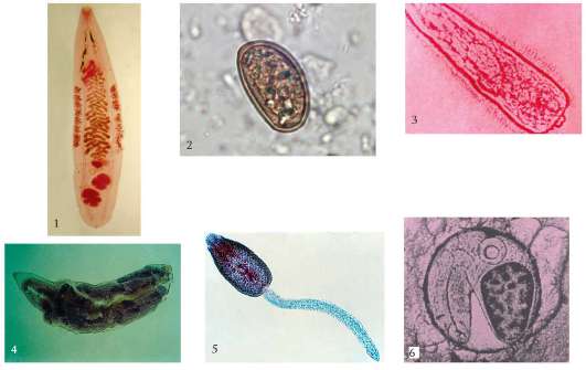
Rice.20.2. Development cycle of flukes: 1 - sexually mature form in the final host; 2 - egg; 3 - miracidium in water; 4 - larva reproducing parthenogenetically in a mollusk; 5 - cercariae in water; 6 - metacercariae in the second intermediate host
In the first case, cercariae either independently invade the host's skin, or, being encystised on plants, turn out to be swallowed by herbivores or humans. In the second case, the cerciaria seek out animals that are used by the main hosts for feeding, and form resting stages in them - encysted metacercariae. Cercariae, unlike miracidia, do not possess chemotaxis, and the use of only geo- and phototaxis, as well as incising on the grass, does not allow them to find specific hosts. Therefore, the bulk of the cercariae die either without finding hosts at all, or after entering the organisms of such species in which development is impossible. After the invasive stages of flukes penetrate into the main host, they migrate in his body and find the organ where they reach puberty and will inhabit the rest of their life.
This means that over the course of evolution, the most perfect mutual adaptations have arisen between flukes and mollusks. Molluscs were probably the first and only hosts of flukes even in the period preceding the emergence of vertebrates. This is also evidenced by the preserved in the development cycle of flukes parthenogenesis- a rudimentary form of sexual reproduction, which takes place in the body of molluscs.
Along with these features, in the developmental cycle of flukes, there are such signs that indicate recapitulation of a free lifestyle: the obligatory release of eggs into the external environment and the presence of actively floating settling stages with developed sensory organs - miracidia and cercariae.
The emergence and further biological and morphophysiological progress of vertebrates, which have mastered all environments favorable for life, opened up wide possibilities of adaptive evolution for flukes by adapting to new hosts. Flukes' adaptations to vertebrates as new habitats were not limited to the use of their wide species diversity. Flukes have adapted to living in a wide variety of organs, tissues and systems - from the skin and sensory organs to the blood vessels of internal organs.
After entering the human body, most flukes carry out complex migrations along the way to the organs of their final localization. Migration occurs through the blood vessels, directly through the spaces between the organs and through the body cavity. During migration, flukes cause severe intoxication and allergic conditions in the host, but it is extremely difficult to diagnose the disease at this moment. Diseases caused by flukes are called trematodes.
Due to the fact that flukes inhabiting humans also infect a number of other mammalian species, the corresponding trematodes are classified as natural focal zoonotic diseases, so their complete elimination is practically impossible.
Developing with one intermediate host and living in the digestive system;
Developing with one intermediate host and living in blood vessels;
Developing with two intermediate hosts.
20.1.1.1. Flukes with one intermediate host inhabiting the digestive system
Herbivores, including domestic animals, become infected much more often than humans. They are the most common source of human infection. Therefore, people in rural areas are usually infected with trematodes of this group.
Laboratory diagnostics- detection of the eggs of these flukes in the feces.
Disease prevention- thorough washing and heat treatment of vegetables and herbs in areas where vegetable gardens are watered with water from stagnant reservoirs, as well as identification, treatment of sick animals and sanitary protection of pastures.
Rice. 20.3.Flukes with one intermediate host: a - hepatic; b - fasciolopsis
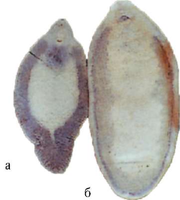
ovaries, vitelline and intestinal branches. The eggs are large, yellowish brown in color. Fascioliasis is more common in countries with warm, humid climates.
Giant liver flukeFasciola gigantica(Fig. 20.3, a) differs from the previous species in large size (up to 75 mm) and weak protrusion of the front of the body forward. The development cycle, diagnosis and prevention do not differ from the previous species. Due to the large size of this type, the fascioliasis caused by it is more difficult. In humans, fascioliasis, caused by a giant liver fluke, is found in Southeast Asia, the Hawaiian Islands, and Uzbekistan.
Diagnostics and prevention fasciolopsidosis correspond to those described above. The disease is widespread in South and Southeast Asia.
20.1.1.2. Flukes with one intermediate host inhabiting blood vessels
This group includes the so-called blood flukes- schistosomes. These are dioecious organisms. Males have a wide body, and females are cord-like and in a sexually mature state are in the gynecophoric canal on the ventral side of males. The suction cups are small and are located at the front end of the body. All blood flukes live in the tropical latitudes of Asia, Africa and America.
As with all flukes, larval stages that reproduce by parthenogenesis develop in aquatic molluscs. Are characteristic cerca-rii- they have a forked tail, and at the front end there are penetration glands, with the help of which they penetrate through the skin into the circulatory system of the final host when it is in water (see Fig. 18.9). However, they cause skin lesions - cercariosis, expressed in the appearance of a rash, itching and other allergic manifestations, as well as in inflammatory reactions. With a massive hit of cercariae in the lungs, pneumonia occurs.
The larvae of schistosomes, pathogenic for humans, migrate through the body and settle in the veins of the abdominal cavity and small pelvis, where they reach puberty. The pathogenic effect of sexually mature schistosomes is expressed in toxic-allergic reactions of the host and local manifestations: bleeding from the affected organs, the formation of ulcers and polyps, prone to malignant degeneration, are characteristic.
Diagnostics consists in the detection of eggs of schistosomes in feces or urine. Allergic intradermal tests and immunobiological reactions are also carried out in vitro.
For the prevention of cercariosis, it is necessary to refrain from swimming in fresh water bodies in which waterfowl live.
Due to the fact that for the penetration of cercariae into human skin it takes at least 15 minutes, it is recommended to quickly wipe the skin with a towel after swimming in such reservoirs.

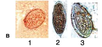
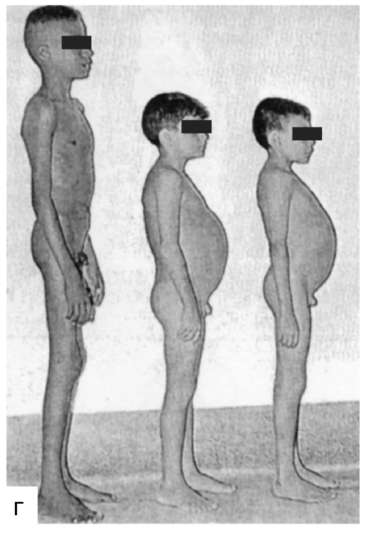
Rice. 20.4.Blood flukes: a - Schistosoma haematobium; b - Schistosoma mansoni; c - eggs with schistosomes: 1 - Sch. japonicum; 2 - Sch. haematobium; 3 - Sch. mansoni; d - children of the same age - on the left - a healthy boy, on the right - growth retardation and puberty in patients with schistosomiasis caused by a fluke Schistosoma mansoni
Schistosoma haematobium- pathogen genitourinary schistosome
per. The male is up to 1.5 cm long, and the female is up to 2.0 cm. The surface of the body is small-knobby. The eggs are very large, up to 0.16 mm in size; there is a long thorn at the end.
Typical for genitourinary schistosomiasis, hematuria (blood in the urine), pain in the suprapubic region, often the formation of stones in the urinary tract. In areas where this disease is spread, bladder cancer occurs 10 times more often than in areas free of schistosomiasis.
When diagnosing find eggs in urine, as well as characteristic changes in the bladder and vagina: ulceration, polypous growths and local inflammatory processes.
In humans, it populates the mesenteric veins of the large intestine and the portal vein system of the liver. In this regard, lesions occur primarily in the large intestine (colitis phenomena, diarrhea mixed with
blood, colon polyposis is possible) and in the liver (venous congestion and cirrhosis). A pronounced delay in physical development is characteristic.
When diagnosing find eggs in feces.
Schistosomes that invade children and adolescents usually lead to a delay in overall physical development and puberty
(Figure 20.4, b).
According to the World Health Organization, about 200 million people are currently affected by various forms of schistosomiasis.
20.1.1.3. Flukes with two intermediate hosts
The ability of cercariae to encyst in the external environment is well known. Probably, in a number of groups of flukes, in the process of evolution, adaptations to encystation in the second intermediate host arose, which increases the likelihood of both survival and getting to the final host. The use of representatives is not only
of different species, but even of different classes and types as such second intermediate hosts (fish, crustaceans, insects) testifies to the independence of the evolution of different groups of flukes in this direction and that this feature of their life cycle has arisen relatively recently. Various directions of the adaptive evolution of flukes of this group led to the fact that they colonized not only different organs of the final hosts, but also different environments, including going on land and losing contact with the original aquatic habitat.
Flukes, which have two intermediate hosts, can be subdivided into those connected in the development cycle with the aquatic environment and not connected, the development cycle of which occurs on land.
20.1.1.3-a. Flukes, whose development cycle is associated with the aquatic environment
Diagnostics based on the detection of eggs in feces.
The second intermediate host is a variety of fish, in which metacercariae are found on scales, fins, gills, and less often in muscles. The circle of final owners is very wide. These are fish-eating birds - pelicans, cormorants, herons - and mammals - minks, otters, bears, as well as humans.
Metagonimus yokogawai(Fig. 20.5, a) - pathogen metagonimosis. This is a small fluke up to 1.5 mm long, its body is densely covered with thorns. The abdominal sucker is located asymmetrically on the right side of the midline. Eggs up to 0.028 mm long.
The first intermediate hosts are gastropods from p. Melania, the second - more than 40 species of fish from this. Carp and Salmon
Rice. 20.5.Flukes with two intermediate hosts: a - Metagonimus yokogawai; b - Nanophyetes salmincola; c - feline fluke; g - Chinese clonorchis; e - pulmonary fluke; e - lanceolate fluke; g - pancreatic fluke
Nanophyetes salmincola(Fig.20.5, b) - pathogen nanofietosis. This fluke has very small dimensions - up to 1.1 mm. The body shape is almost round. The eggs are relatively large, up to 0.056 mm long. The first intermediate host is mollusks from the r. Semisulcospira, the second - this fish. Salmon, Grayling, Carp and Podkamenschik. The final hosts are fish-eating mammals and humans. It is found in the Amur River basin in the Primorsky Territory, on Sakhalin and on the western coast of North America. Human diseases have been described only in the local population of the middle and lower reaches of the Amur River.
20.1.1.3-c. Flukes living in the bile ducts of the liver
Feline flukeOpisthorchis felineus(Fig. 20.5, c) - pathogen opisthorchiasis. Body length up to 13 mm. A characteristic feature is two well-stained lobe-shaped testes at the posterior end of the body. Eggs are 26-30 microns long, with a lid. Opisthorchiasis is an endemic disease in Russia. In humans, it is most often found in Western Siberia, but occasionally manifests itself in the European part of the CIS - in the Volga-Kama basin, in the basin of the Don, Dnieper, Dniester and Seversky Donets rivers. Found in the Nemunas basin. Natural foci without human participation are also known in Kazakhstan.
Opisthorchis viverrini- pathogen civet opisthorchiasis. It differs from the previous species in large-lobed testes, small body size (up to 10 mm) and distribution area. This is a typical tro-
pike worms, common in Thailand, Laos and Malaysia, where in some areas the infection rate of the population reaches 90%. As in the previous species, the hosts are mollusks from p. Bithynia and carp fish. The final hosts are primarily carnivorous mammals from this. Viverrids, rarely cats, dogs and humans.
Clonorchis sinensis(Fig.20.5, d) - pathogen clonorchiasis. This fluke is larger than the previous two - up to 25 mm in length. The shape of the testes is characteristic: they are branched and are located one after the other in the back of the body. Eggs up to 30 microns long. Distributed in Southeast Asia, in the Far East. In Russia - in the south of the Primorsky and Khabarovsk Territories. The first intermediate hosts are mollusks of the genera Bithynia and Parafossularis, the latter - more than 70 species of carp fish, less often goby and herring. The final hosts are humans and fish-eating mammals.
20.1.1.3-d. The flukes living in the lungs
a person's life outside these regions is random, sporadic. The persistence of national traditions in nutrition, as well as the emphasized natural focal nature of paragonimiasis, complicate the prevention of this disease.
Lung flukes have an unusual body shape for flukes: they resemble orange seeds and are up to 12 mm in size. Eggs up to 0.118 mm long.
The first intermediate host is mollusks from the genera Semisulcospira, Oncomelania and some others. The second intermediate host is crabs from the genus Eriocheir, Potamon, childbirth cancers Cambarus, Procambarus, as well as shrimp p. Macrobrachium. The final owners are humans and animals that feed on crustaceans - otters, minks, pigs, cats, dogs and some rodents.
For diagnostics diseases, it is necessary to examine the sputum of patients, in which eggs are found, as well as feces, where eggs can get when the sputum is swallowed. They do not change in the digestive tract.
Personal prevention paragonimiasis consists in the refusal to eat crustaceans that have not undergone heat treatment. Community prevention corresponds to measures applied against flukes developing in an aquatic environment with two intermediate hosts.
20.1.1.3-d. Flukes, whose development cycle is not associated with the aquatic environment
The first intermediate host is mollusks of the genera Helicella or Zebrina, the second is the ant p. Formica. A person becomes infected by accident by swallowing an infested ant. This fluke settles in the bile ducts of the liver.
Diagnostics- as with all trematodes with liver damage.
Personal prevention- it is necessary to ensure that ants do not get into food. Public prevention - deworming of livestock and sanitary protection of pastures.
Diagnostics- detection of eggs in feces and duodenal contents during probing.
Prophylaxis human infection - refusal to eat insects or their preliminary heat treatment. For the rest, see the previous view.
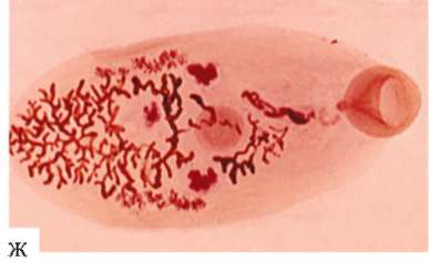
20.1.2. TAPE CLASS CESTOIDEA
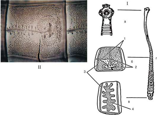
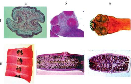
Rice. 20.6.I - organization of tapeworms: a - head; b - hermaphroditic segment; c - a mature segment; g - strobe; 1 - male genital organs; 2 - female genital organs; 3 - channels of the excretory system; 4 - uterus with mature eggs; II - hermaphroditic segment of the bovine tapeworm
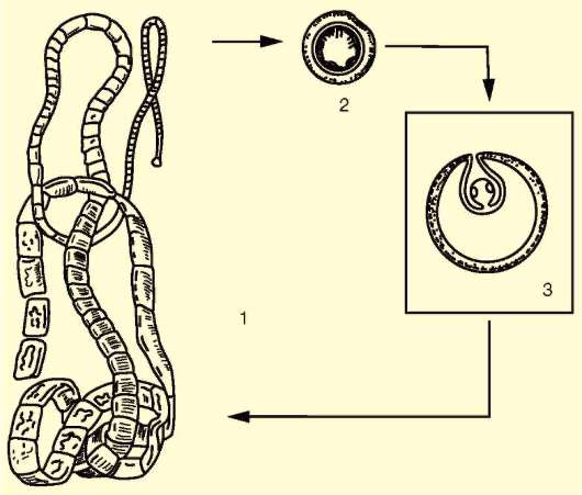
Rice. 20.7.The developmental cycle of tapeworms: 1 - sexually mature stage in the intestine of the final host; 2 - an egg in the external environment; 3 - the finnose stage in the tissues of the intermediate host
If the main host, in whose intestines the helminth is located, suffers from it relatively slightly, then the viability of the intermediate host with the Finns of these worms in the lungs, brain or liver is sharply reduced. This increases the likelihood that it is the infested animals that will be eaten by the final host.
According to the characteristics of biology, tapeworms of medical importance can be divided into groups, the life cycle of which is associated and not associated with the aquatic environment. The second group is subdivided into helminths:
Using man as the ultimate master
Dwelling in a person as in an intermediate master,
The entire life cycle takes place in a person.
Diseases caused by tapeworms are called cestodosis. Many types of tapeworms infect only humans, others are also found in natural settings, they are characterized by the existence of classical natural foci. Cestodes, living in humans as the main host, are brought together by the fact that they live in the intestines and are always in small numbers. This is due to pronounced intraspecific competition, in which only a few individuals survive. This does not affect the intensity of reproduction, since their fertility is enormous.
20.1.2.1. Tapeworms whose life cycle is associated with the aquatic environment
two intermediate hosts living in the aquatic environment - small planktonic crustaceans from the genera Cyclops and Diaptomus, as well as fish that feed on them. The crustaceans are inhabited by a larva called procercoid, in fish - plerocercoid. A primitive feature of the plerocercoid is its ability to actively move, and in the case of eating smaller fish invaded by plerocercoids, the latter pierce the intestinal wall and enter the abdominal cavity and muscles. Thus, large fish can accumulate hundreds of plerocercoids throughout their life. The host becomes infected by eating the invasive fish. Therefore, the basis for personal prophylaxis is the thermal treatment of fish products.
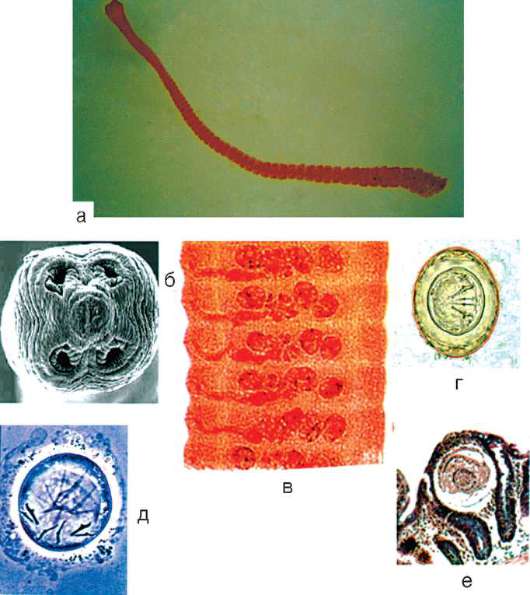
Wide ribbonDiphyllobothrium latum(Fig. 20.8, a) is the most common causative agent of diphyllobothriasis in humans. The strobila has a length of about 10 m. The head is equipped with bothria- suction gaps, mature segments are characterized by a small rosette uterus. The uterus has a connection with the external environment, therefore, ripening eggs are freely removed from it. Eggs are yellowish in color, up to 71 microns long, have a lid on one pole, and on
Rice. 20.8.Large tapeworms: I - heads; II - mature segments: a - wide tapeworm (cross section): 1 - cross section; b - bovine tapeworm (general view); c - pork tapeworm (general view)
Personal prevention infection - see above. Community prevention- protection of reservoirs from fecal contamination.
diagnostics of diseases do not differ from those given for the broad tapeworm.
20.1.2.2. Tapeworms whose life cycle is not associated with the aquatic environment
20.1.2.2-a. Tapeworms using humans as their ultimate host
Personal prevention- heat treatment of meat. Community prevention- sanitary control of meat products and sanitary and educational work with the population.
Bovine tapewormTaeniarrhynchus saginatus(Fig.20.8, b) - pathogen teniarinchosis, reaches a length of 4-10 m. It has only four suction cups on the head. The hermaphroditic segments are square in shape, the uterus does not branch in them, and the ovary consists of two lobes. Mature segments are strongly elongated. The uterus is very ramified, the number of its lateral branches reaches 17-34 pairs. Eggs contain oncospheres located under a thin transparent shell, which quickly disintegrates. Oncospheres have three pairs of hooks and a thick, radially striated
loch. The diameter of the oncospheres is about 10 microns. Teniarinhoz is widespread everywhere, where the population eats raw or insufficiently processed beef meat.
Diagnostics simple - when mature segments are found in feces, since the segments have a characteristic structure.
Prophylaxis teniarhynchosis is to protect pastures from contamination with human feces.
Intermediate hosts of this helminth, in addition to domestic and wild pigs, can be cats, dogs and humans. In this case, they, like pigs, develop cysticercosis. A person can swallow pork tapeworm eggs by accident, but more often cysticercosis occurs as a complication of teniasis. In this disease, reverse intestinal motility and vomiting are especially common. Mature segments can thus enter the stomach, be digested there, and the released oncospheres penetrate the intestinal vessels, are carried by blood and lymph throughout the body, where in the liver, muscles, lungs, brain and other organs
cysticercus is formed. This can lead to rapid death.
Laboratory diagnostics teniasis is based on the detection of characteristic mature segments in feces; the diagnosis of cysticercosis is more difficult - by means of X-ray examination and staging of immunological reactions.
For personal prevention teniasis must be thermally treated pork, and cysticercosis must follow the rules of personal hygiene. Community prevention- closed keeping of pigs.
Table 20.1. Tapeworms accidentally using humans as their ultimate host
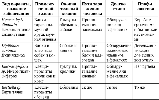
20.1.2.2-6. Tapeworms using humans as an intermediate host
Laboratory diagnostics larval cestodoses is complicated by the fact that the Finns have no connection with the environment and, apart from dissimilation products, do not emit anything. The diagnosis is made on the basis of X-ray, biochemical and immunological studies.
EchinococcusEchinococcus granulosus(Fig. 20.9, a) - pathogen echi nococcosis. The sexually mature form has a head with hooks and 3-4 segments of varying degrees of maturity. The last one is mature and contains about 800 eggs. The total body length is up to 5 mm. The eggs are similar in shape and size to those of pork and bovine tapeworms. Echinococcosis in humans is widespread in all geographical and climatic zones, mainly in regions with developed distant-pasture animal husbandry.
Life cycle echinococcus is associated with predatory animals of the Canidae family (wolves, jackals, dogs), which are its final owners. Adult segments are able to actively reproduce
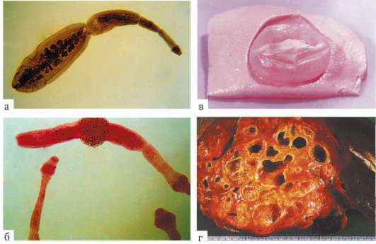
Rice. 20.9.Tapeworms using humans as an intermediate host: a - echinococcus; b - alveococcus; c - a single Finna of an echinococcus in the liver; d - budding finna alveococcus in the liver
nest, spreading eggs through the owner's coat and in the environment. They can be swallowed by herbivores - cows, sheep, deer or humans, becoming intermediate hosts. Finna echinococcus is a bubble, often reaching 20 cm in diameter. It is filled with liquid with a huge number of young scolexes constantly budding from the inner surface of the Finnish wall. The final host becomes infected by eating the affected organs of the intermediate.
The growing Finn compresses the organs, causes them to atrophy. The constant supply of dissimilation products into the host's organism causes its depletion. The rupture of the echinococcal bladder is very dangerous: the liquid contained in it can cause toxic shock. In this case, small embryonic scolexes can spread throughout the body, affecting other organs. Multiple echinococcosis usually ends with the death of the host.
Personal prevention infection - washing hands after contact with herding dogs. Community prevention- examination and deworming of dogs, prevention of feeding them organs of sick animals.
Alveococcosis is a more serious disease than echinococcosis, due to the invasive nature of the growth of the Finns, from which it is possible for small daughter Finns to bud and settle them through the lymphatic system of the host into different organs.
Personal prevention- as with echinococcosis, public- observance of the rules of hygiene when processing the skins of game animals, as well as the prohibition of feeding rodent carcasses to dogs.
The described type of helminth is settled in East and Southeast Asia, Australia, America and Africa.
Diagnostics complicated. Usually, the diagnosis is made during surgery.
Prophylaxis- filtration of water used for drinking, and heat treatment of exotic food products - meat of frogs and snakes.
Sparganum proliferum(Fig. 20.10, b) - plerocercoid of an unknown species of tapeworm, probably also from p. Spirometra. It is characterized by an original feature - the ability to bud like an alveococcus, but forms no more than several daughter individuals, morphologically related to the mother. In this regard, it received the name proliferum- growing. Its dimensions coincide with the dimensions of the previous type. The life cycle of this species is poorly understood; it probably corresponds to the development of the previous species; helminth is most often found in Korea, Vietnam and Japan. In addition to the methods of infection characteristic of the previous species, Sparganum can penetrate
into the human body through ulcerated skin and mucous membranes when applied to them as a folk oriental remedy for snake meat containing plerocercoids.
20.1.2.3. Tapeworms that go through the entire life cycle in the human body
Hymenolepiasis is ubiquitous, especially in countries with dry and hot climates. Mostly children are affected.
Rice. 20.11.Dwarf tapewormHymenolepis nana:a - general view; b - head; c - segments; g - egg; e - oncosphere; e - cysticercoid in the villus of the human intestine
For diagnostics detection of eggs in feces is important.
20.2. TYPE ROUND WORMNEMA THELMINTHES
The body shape of these organisms is elongated fusiform or filamentous. The musculocutaneous sac consists of a multilayer, dense, elastic and inextensible cuticle, hypodermis, which is a single cytoplasmic mass, not divided into individual cells and containing a large number of nuclei, and one layer of longitudinal smooth muscles. The cuticle is mainly protective. The muscles are located in the form of two longitudinal strands - on the dorsal and ventral sides of the body. Their alternate contraction provides vigorous flexion and extension movements and rapid movement of the body in space.
The digestive system is in the form of a through tube with mouth and anus. The nervous system is represented by longitudinal cords connected by annular bridges. The excretory system is based on a protonephridial structure, but the number of excretory cells is counted in units. Roundworms are dioecious. The reproductive system is built in the form of tubes differentiated along the length, some of which function as ovaries or testes, some as vas deferens or oviducts, and some as organs, in
which accumulate and retain mature reproductive products. All internal organs are located in the primary body cavity, filled with fluid, which gives the entire body elasticity and ensures the exchange of substances between organs (Fig. 20.12).
A peculiar feature of round worms is that a certain number of cells are always included in their body. This limits their ability to grow and regenerate. Only representatives of the class Actually roundworms are of medical importance.
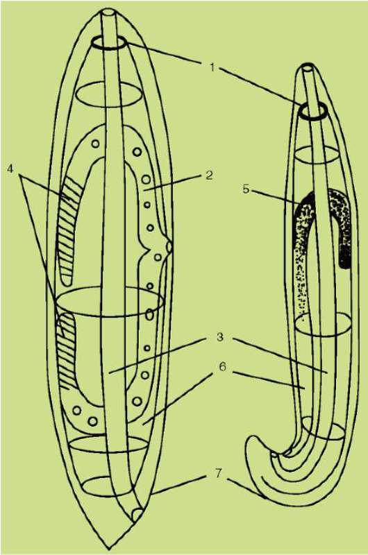
Rice. 20.12.Organization of roundworms: a - female; b - male: 1 - nervous system; 2 - uterus; 3 - intestines; 4 - ovaries; 5 - testis; 6 - primary body cavity; 7 - cuticle
20.2.1. CLASS PROPERLY ROUND WORM NEMATODA
20.2.1.1. Roundworms - geohelminths
In these worms, eggs or larvae necessarily develop in the surface layers of the soil when oxygen is available and sufficient moisture is available. With the exception of pinworms, all helminths of this group are found more often in regions with hot and humid climates, which provide larvae and eggs with more opportunities to develop in the soil. Geohelminths are not found in the Arctic and southern circumpolar regions. Males and females are easily distinguishable: males of most species have a bent to the ventral side or a spirally twisted posterior end of the body, while in females it is straight.
For diagnostics of all nematodes of this group, it is important to detect eggs in the patient's feces.
Preventive measures are aimed at preventing the ingress of invasive eggs into the digestive system - personal hygiene and food hygiene, less often - other measures. Some of the geohelminths, entering the human digestive system, quickly reach puberty and begin to multiply in the intestines without migrating through the host's body. The larvae of others, before reaching puberty, necessarily move through the blood vessels and the respiratory system and only then develop in the intestines.
20.2.1.1-a. Geohelminths developing without migration

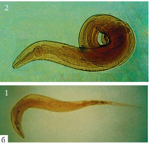
Rice. 20.13.Geohelminths developing without migration: a - whipworm; b - pinworm: 1 - females; 2 - males
Trichocephalosis is very widespread, but most often occurs in countries with subtropical and tropical climates. It currently affects about 500 million people.
Children's pinwormEnterobius vermicularis(Fig.20.13, b) - pathogen enterobiasis- the most common helminthiasis, which occurs mainly in children. Small worm with a pronounced sex
dimorphism: the female reaches a length of 12 mm, her body is straight, pointed behind. Males are smaller (up to 5 mm) and coiled spirally. Eggs are oval, somewhat asymmetrical, transparent and colorless, up to 50 microns in length.
Laboratory diagnostics has features: eggs are rarely found in feces, so they are examined in a smear from the perianal folds of the perineum.
Personal prevention- observance of the rules of personal hygiene, especially thorough washing of hands after sleep, short cutting of nails. Public- regular examination of children in childcare facilities, treatment of patients, regular examinations of workers in childcare facilities and public catering establishments.
20.2.1.1-6. Geohelminths developing with migration
The peculiarity of this group of geohelminths is that, having entered the human body by any means, at the larval stage they first penetrate the veins of the systemic circulation, then enter the heart and from there into the small circle. In the capillaries of the lungs, they leave the bloodstream into the alveoli, actively rise along the bronchi, trachea, and larynx. These movements cause a cough reflex, which promotes their exit into the pharynx and secondary swallowing. Only after that, entering the intestines, the larvae reach sexual maturity.
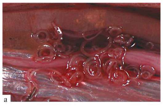
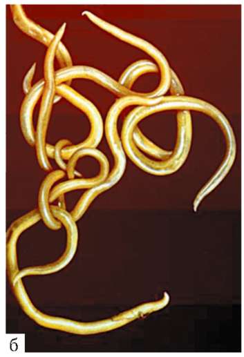
Rice. 20.14.Ascaris human: a - female and male; b - larvae penetrating the intestinal wall; c - mature roundworm egg
Ascaris humanAscaris lumbricoides(fig.20.14) - pathogen ascariasis- a large helminth, females of which reach 40 cm in length, and males - 20 cm. Mature eggs are oval and tuberous, their shell is thick and multilayered. The color is yellowish brown, length up to 60 microns. The fertility of the roundworm is enormous: the female lays up to 20,000 eggs daily.
This species is very close to the pig roundworm, which in Southeast Asia can easily infect humans and, conversely, humans - pigs. This indicates their relatively recent origin from the same ancestor and the time of the beginning of the divergence not earlier than the beginning of the domestication of pigs, which took place in Southeast Asia.
Life cycle typical for helminths of this group. Eggs ripen at high soil moisture and a temperature of 18-25 ° C in 2-3 weeks. Invasion of large numbers of ascaris can lead to blockage of the intestine or common bile duct. There are known cases of atypical localization of ascaris: in the larynx, middle ear, liver and even the heart. This requires urgent surgical intervention.
According to the WHO, 700 million people are currently suffering from ascariasis.
Krivogolovka duodenumAnkylostoma duodenale and nekatorNecator americanus(fig. 20.15) - both types belong to this. Ankylostomids.
Rice. 20.15.Krivogolovka of the duodenum: a - sexually mature male; b - egg; c - the larva emerging from the egg
Life cycle hookworm is peculiar and testifies to their close connection with free-living ancestral forms. Eggs that enter the soil develop rapidly, and larvae soon emerge from them, which, after molting twice, after a few months become
invasive and can enter the human body either with soil-contaminated vegetables and fruits, or through active penetration through the skin.
Laboratory diagnostics- as with all geohelminthiasis.
Prophylaxis- in addition to the general measures described above, the mandatory wearing of shoes in areas where these diseases are common.
Ankylostomiasis currently affects 700 to 900 million people worldwide.
Like hookworms, intestinal acne is found mainly in the tropical and subtropical zones. It is especially widely settled in Southeast Asia, South and Central America. The existence of underground foci in zones with a temperate climate is possible, as in ankylostomiasis.
When diagnosing a reliable sign of the disease is the detection of live larvae in feces, vomit and during duodenal intubation.
Prophylaxis strongyloidosis corresponds to the prevention of ankylostomiasis.
20.2.1.2. Roundworms - biohelminths
20.2.1.2-a. Biohelminths, the infection of which occurs when the larvae are swallowed with the tissues of the intermediate host
These helminths are characterized by primary localization in the human intestine, and then penetration into the blood through its wall and further into the tissues of the internal environment. Prevention in this regard is the creation of obstacles to the penetration of larvae through the mouth.
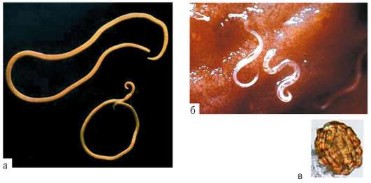
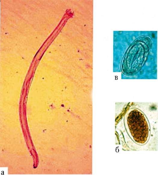
Guinea worm Dracunculus medinensis(fig.20.16) - pathogen dragon-leza. The length of the female is up to 120 cm, the male is only 2 cm.
The disease is widespread in zones with tropical and subtropical climates, previously it was found in Central Asia.
Personal prevention it is also simple - boiling or filtering drinking water taken from open reservoirs. Community prevention- modern water supply with disinfected water; identifying and treating patients guarantees success in the fight against this disease.
Dracunculiasis is now considered a vanishing disease, although 50 years ago it affected tens of millions of people, mostly in North and Central Africa, Pakistan and India.
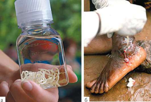
A person becomes infected by eating the meat of infected animals, most often pigs. The swallowed larvae reach puberty quickly in the intestines. Each fertilized female gives birth to about 1,500 live larvae, after which she dies. Larvae bore through the wall
Rice. 20.17.Trichinella spiral: a - Trichinella larvae encapsulated in muscles; b - sexually mature individuals from the human intestine: 1 - female; 2 - male
intestines and, transported by blood, settle in striated muscles: most often in the diaphragm, intercostal, deltoid. Here, after the destruction of a part of the muscle fibers, they are spirally twisted and encapsulated.
eating not only rats and mice, but also insects, worms, fish, corpses and bird droppings, in which viable larvae are preserved.
The pathogenic effect of Trichinella includes both general allergic phenomena and muscle dysfunction.
Diagnosis based on history data - the use of wild meat and pork not verified by the veterinary service, as well as on the results of muscle biopsy.
Personal prevention- thorough heat treatment of pork and especially meat of wild animals. Community prevention- sanitary supervision in pig breeding, inspection of pork at retail outlets and at public catering establishments.
20.2.1.2-6. Biohelminths transmitted by transmission
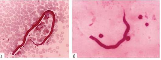
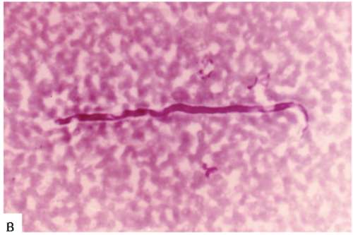
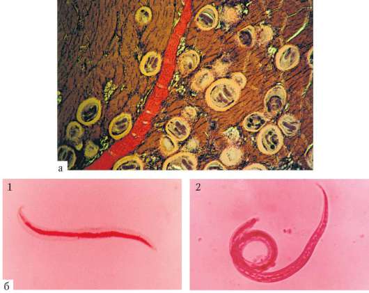
Rice. 20.18.Microfilariae from human blood: a - Wuchereria bancrofti; b - Onchocerca volvulus; v - Brughia malayi
hours. In the case when biting midges serve as carriers, the activity of which depends more on air humidity, the release of microfilariae into the blood is devoid of periodicity. These features of the biology of filariae must be taken into account when making a diagnosis and take blood from patients to detect microfilariae in it at a time when their presence there is most likely. Prophylaxis filariasis - identification and treatment of patients, vector control (see clause 21.2.2).
Currently, about 150 million people are infected with various forms of filariasis.
20.2.1.3. Roundworms that carry out only migration in the human body
Prophylaxis- hygienic keeping of pets.
The pathogenic effect is associated with a violation of the integrity of the stomach wall, which is accompanied by severe pain and gastritis. Possible perforation of the stomach and peritonitis, ending with the death of the patient.
The disease is widespread in the countries of the Far Eastern region, especially well known in Japan, where seafood is usually eaten raw.
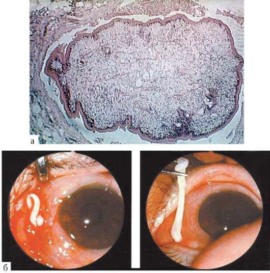
Diagnosis set during gastroscopy, when larvae are found that have invaded the gastric mucosa (Fig. 20.19, b).
Rice. 20.19. Larva Anisakis sp .: a - in the liver of fish; b - from the human stomach
Prophylaxis anizakiaza - sanitary control of sea products, their heat treatment during cooking.
Questions for self-control
1. Characteristics and classification of the type Flatworms.
4. Can a person be an intermediate master of flukes?
5. Can humans be an intermediate host for tapeworms?
6. Characteristic of the Roundworm type.
7. Roundworms geo- and biohelminths. Features of life cycles, ways of infection and preventive measures.
8. Are fascioliasis, teniasis, alveococcosis, ankylostomiasis, ascariasis, trichinosis, enterobiasis and noncatorosis natural focal diseases?