1. What are the features external structure hydra?
Hydra is an elongated sac-shaped polyp, reaching 1.5 cm in length. It is attached to the substrate by a sole located at one end of the body. At the other end there is a mouth opening surrounded by a corolla of tentacles. The hydra body wall is formed by two layers of cells: the outer - ectoderm and the inner - endoderm.
2. How is the ectoderm of coelenterates organized?
Several types of cells can be distinguished in the ectoderm. The bulk is represented by epithelial-muscle cells that have processes in which contractile elements are concentrated. Also in the ectoderm are sensory, nervous, glandular, stinging and intermediate cells. Sensitive cells are located in the same way as epithelial-muscular cells, i.e., one end faces outward and the other is adjacent to the basement membrane. Nerve cells lie between the contractile processes on the basement membrane. Intermediate cells are undifferentiated cells from which specialized cells subsequently develop; in addition, they participate in regeneration. Sex cells are formed in the ectoderm.
3. What type of nervous system do coelenterates have?
Coelenterates have a diffuse type of nervous system. Sensitive cells are located in the same way as epithelial-muscular cells, i.e., one end faces outward and the other is adjacent to the basement membrane. Nerve cells lie between the contractile processes on the basement membrane. If you touch the hydra, the excitation that arises in the primary cells quickly spreads throughout the entire nervous network and the animal responds to the irritation by contracting the processes of the epithelial-muscular cells.
4. How does a hydra stinging cell work?
The largest number of stinging cells are located in the tentacles. Inside the cell there is a stinging capsule with a poisonous liquid and a spirally coiled hollow thread. On the surface of the cell there is a sensitive spine that perceives external influences. In response to irritation, the stinging capsule throws out the thread it contains, which turns out like the finger of a glove. Burning or poisonous contents are released along with the thread. Thus, hydroids can immobilize and paralyze fairly large prey, such as cyclops or daphnia. Stinging cells are replaced with new ones after use.
5. What cells form the inner layer of the hydra?
The cellular elements of the endoderm are represented by epithelial-muscular and glandular cells. Epithelial muscle cells often have flagella and processes resembling pseudopodia. Glandular cells secrete digestive enzymes into the digestive cavity: greatest number such cells are located near the mouth.
6. Tell us about the nutrition of hydra.
Hydra is a predator. It feeds on plankton - ciliates, small crustaceans (cyclops and daphnia). The stinging threads entangle the prey and paralyze it. Then the hydra grabs it with its tentacles and directs it into the mouth opening.
7. How does the hydra digestion process?
Digestion in hydras is combined (intracavitary and intracellular). Swallowed food enters the digestive cavity. First, the food is processed with enzymes and crushed in the digestive cavity. Then food particles are phagocytosed by epithelial-muscle cells and digested in them. Nutrients diffusely distributed among all cells of the body. From the cells, metabolic products are released into the digestive cavity, from where, along with undigested food debris, they are thrown into the digestive cavity. environment through the mouth opening.
8, What are intermediate cells, what are their functions?
Intermediate cells are undifferentiated cells that give rise to all other types of ecto- and endoderm cells. These cells ensure the restoration of body parts when damaged - regeneration.
9. What is hermaphroditism?
Hermaphroditism is the simultaneous presence of both male and female organs in one organism (from the Greek Hermaphroditos - the son of Hermes and Aphrodite, a mythical bisexual creature).
10. How does hydra reproduce and develop?
Hydra reproduces asexually and sexually.
During asexual reproduction, which occurs during a period favorable for life, one or more buds are formed on the body of the mother’s body, which grow, their mouth breaks through and tentacles form. Daughter individuals are separated from the mother. Hydras do not form real colonies.
Sexual reproduction occurs in autumn. Hydras are mostly dioecious, but there are also hermaphrodites. Sex cells are formed in the ectoderm. In these places, the ectoderm swells in the form of tubercles, in which either numerous spermatozoa or one amoeboid egg are formed. Spermatozoa, equipped with flagella, are released into the environment and delivered to the eggs by a current of water. After fertilization, the zygote forms a shell, turning into an egg. The maternal organism dies, and the shell-covered egg overwinters and begins development in the spring. The embryonic period includes two stages: cleavage and gastrulation. After this, the young hydra leaves the egg shells and goes outside.
11. What are hydromedusae?
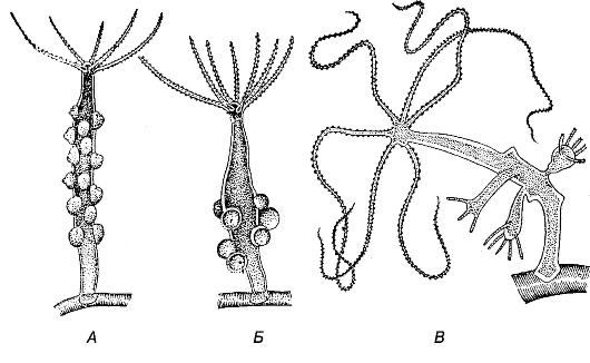
Hydromedusae are free-swimming sexual specimens of some representatives of the hydroid class; they are formed by budding.
12. What is planula?
Planula is a larva covered with cilia. Formed after fertilization in some hydroids. Attaches to underwater objects and gives rise to a new polyp.
13. What does it feel like internal structure coral polyp?
Coral polyps have all the characteristic features of coelenterates.
The body of coral polyps has the shape of a cylinder. They have a mouth surrounded by tentacles leading into a throat. The digestive cavity is divided into a large number of chambers, thereby increasing its surface and, consequently, the efficiency of food digestion. There are muscle fibers in the ecto- and endoderm that allow the polyp to change its body shape.
A characteristic feature of coral polyps is that most of them have a hard calcareous skeleton or a skeleton consisting of a horn-like substance.
14. What role do coelenterates play in nature?
Coelenterates are predators and occupy a corresponding niche in food chains reservoirs, seas and oceans, regulating the number of single-celled, small crustaceans, worms, etc. Some deep-sea species of jellyfish feed on dead organisms.
Coral polyps, which live in shallow tropical seas, form the basis of reefs, atolls and islands. These corals play an important role in coastal communities that include significant numbers of animals and plants.
TYPE COELENTERATATYPE COELENTERATA
Hydroid class (Hydrozoa)
Hydra freshwater (Hydra fusca)
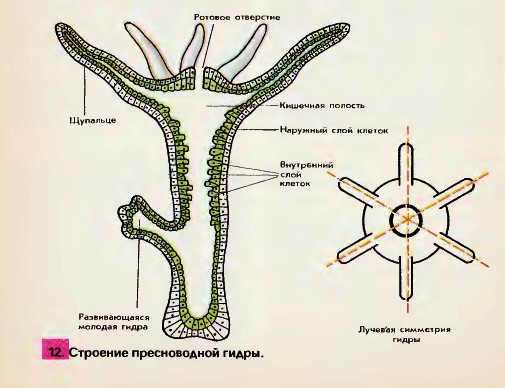
A representative of hydroids is freshwater hydra. Hydra lives in ponds, lakes, rivers, and has a cylindrical shape. At one end there is a mouth surrounded by 5 - 12 thin long tentacles, on another - sole. Using the sole, the hydra is attached to objects. The hydra's body is 1 - 1.5 cm long.
Hydra is characterized by radial symmetry(Fig. 94).
The walls of the hydra's body consist of two layers: the outer - ectoderm and internal - endoderm. Between them there is a structureless mass - mesoglea.
Inside the hydra's body is gastric cavity.
The mouth opening serves for eating and removing undigested residues (Fig. 95).
Rice. 94.Longitudinal section of a freshwater hydra.
Ectoderm cells are differentiated into epithelial, epithelial-muscular, interstitial (intermediate), stinging, nervous.
Epithelial muscle cells have a body and two contractile processes. These processes are located along the body. When they contract, the body thickens and shortens.
Between the epithelial-muscle cells are small interstitial cells. Due to them, reproductive and stinging cells are formed. The stinging cells contain an oval stinging capsule with dense walls. The capsule is filled with liquid; Inside the capsule there is a spiral thread, on the surface of the cell there is a thin tactile hair. When this hair is irritated, the stinging capsule throws out an elastic thread. The stinging cells serve the hydra for attack and defense (Fig. 96).
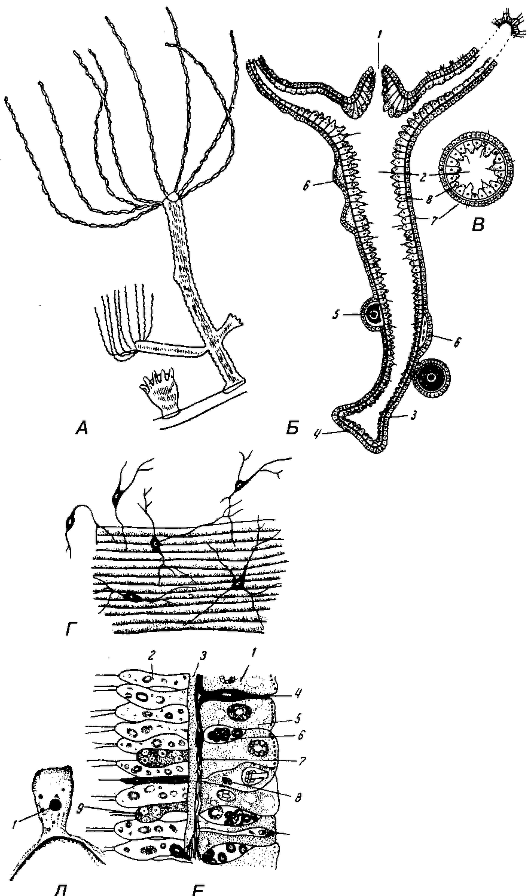
Rice. 95. Hydra Hydra fusca.
A- general form hydra; B- lengthwise cut: 1 - mouth; 2 - gastric cavity; 3 - stalk; 4 - sole; 5 - egg cell; 6 - spermatozoa: 7 - ectoderm; 8 - endoderm; IN- cross section; G - nerve cells; D - ectodermal epithelial-muscle cell: 1 - core; E - longitudinal section of the hydra body wall: 1 - ectoderm cell; 2 - endoderm cell; 3 - mesoglea; 4 - nerve cell; 5 - epithelial muscle cell; 6 - interstitial cell; 7 - basement membrane; 8 - stinging cell; 9 - glandular cell.
Rice. 96.Stinging cell. 1 - stinging capsule; 2 - tactile hair; 3 - stinging thread with spines; 4 - spikes; 5 - core.
Rice. 97.The location of nerve cells in the body of the hydra (according to Hesse).
Rice. 98.Hydra irritability.
The ectoderm contains star-shaped nerve cells. They are connected to each other by processes, forming a diffuse nervous system (Fig. 95 (d), 97, 98).
Endodermlines the entire gastric cavity (Fig. 99).
Rice. 99.The structure of the endoderm cell (inner layer) of the hydra body.
Cells endoderm differentiated by epithelial-muscular, digestive, glandular, nervous.
The muscular processes of endodermal epithelial-muscular cells are located transversely with respect to the longitudinal axis of the body. When they contract, the hydra's body narrows and becomes thinner.
The epithelial part of the endodermal cells, directed towards the gastric cavity, bears 1-3 flagella and is capable of forming pseudopodia, which can capture small food particles. This is intracellular digestion.
The glandular cells of the endoderm secrete digestive juices directly into the gastric cavity, where digestion also occurs. Hydra combines intracellular and cavity digestion. Hydra feeds on daphnia and cyclops. Hydra breathes over the entire surface of its body.
Hydra breeds asexual And sexually(Fig. 100).
At asexual reproduction on the body of the hydra are formed kidneys They gradually increase in size, take the shape of a hydra and separate from the mother’s body (Fig. 101).
Rice. 100.Hydra fusca at low magnification.
A- hydra with male gonads; B- hydra with female gonads; IN-
budding hydra (according to Polyansky).
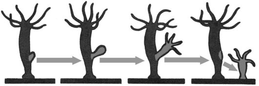
Rice. 101.Hydra budding.
When the temperature drops, hydra reproduces sexually.
Hydra - hermaphrodite. Germ cells are formed from interstitial cells of the ectoderm. Tubercles appear at the sites where germ cells are formed.
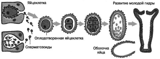
Rice. 102.Hydra sexual reproduction.
The eggs are located closer to the base, and the sperm are located closer to the mouth of the hydra. Cross fertilization. In autumn, the egg is fertilized in the mother's body, surrounded by a dense shell, then the hydra dies. The eggs remain dormant until spring, when new hydras develop from them (Fig. 102).
Hydra is capable of regeneration.
Questions for self-control
1. What organisms are multicellular?
2. Who is a representative of the hydroid class?
3. Where does hydra live?
4. What is the structure of hydra?
5. How many layers of cells does the hydra’s body consist of?
6. How are ectoderm cells differentiated?
7. What structure do stinging cells have and what functions do they perform?
8. How are endoderm cells differentiated?
9. How does hydra digestion? 10.How does hydra reproduce?
11.How it happens asexual reproduction at the hydra? 12. How it happens sexual reproduction at the hydra?
Key words of the topic “Subkingdom Multicellular. Type Coelenterates"
tubercles spring
intracellular digestion
performance
gastric cavity
hermaphrodite
hydra
hydroid daphnia
differentiation diffuse nervous system flagella
glandular cells animals liquid protection
star shape
coelenterates
cells
interstitial cells
nerve cells
stinging cells
epithelial muscle cells
epithelial cells
maternal organism
mesoglea
multicellular
attack
undigested remains
education
lake
fertilization
organism
organs
autumn
base
tactile hair relation
digestive juices
food particles
surface
sole
sub-kingdom
item
representative
meal
process
pond
pseudopodia work
radial symmetry
irritation
dimensions
asexual reproduction
sexual reproduction
regeneration
result
rivers
genus
mouth opening
system
layers
contractile processes resting state spiral filament body wall stinging capsule body tissue type
elastic thread function
functional unit
Cyclops
tentacles
endoderm
>>Freshwater polyp hydra
Multicellular animals
Type coelenterates
Coelenterates are bilayered multicellular animals with a single body cavity - the intestinal cavity (hence the name of the type).
§ 7 Freshwater polyp hydra
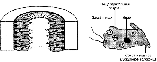
Habitat, structural features and life activity. In lakes, rivers or ponds with clean, transparent water, a small translucent animal - a polyp - is found on the stems of aquatic plants. hydra(“polyp” means “multi-legged”). It is an attached or sedentary coelenterate with numerous tentacles 12 . The body of an ordinary hydra has an almost regular cylindrical shape. At one end there is a mouth surrounded by a corolla of 5-12 thin long tentacles, the other end is extended in the form of a stalk with a sole at the end. Using the sole, the hydra is attached to various underwater objects. The body of the hydra, together with the stalk, is usually up to 7 mm long, but the tentacles can extend several centimeters.
Radiation symmetry. If you draw an imaginary axis along the body of the hydra, then its tentacles will diverge from this axis in all directions, like rays from a light source. Hanging down from some aquatic plant, the hydra constantly sways and slowly moves its tentacles, lying in wait for prey. Since the prey can appear from any direction, the tentacles arranged in a radial manner are best suited to this method of hunting.
Radiation symmetry is characteristic, as a rule, of animals leading an attached lifestyle.
1 - hydra; 2 - wild boar; 3 - mouse; 4 - butterfly; 5 - hawk on the nest; 6 - dolphins in the dolphinarium.
Intestinal cavity. The body of the hydra has the form of a sac, the walls of which consist of two layers of cells - the outer (ectoderm) and the inner (endoderm). Inside the hydra's body there is an intestinal cavity (hence the name of the type - coelenterates).
The outer layer of cells is the ectoderm. Under a microscope, several types of cells are visible in the outer layer of the hydra - the ectoderm. 13 . Most of all here are skin-muscular. By touching their sides, these cells create the cover of the hydra. At the base of each such cell there is a contractile muscle fiber, which plays an important role in the movement of the animal. When the fibers of all skin-muscle cells contract, the hydra's body contracts. If the fibers contract on only one side of the body, then the hydra bends in that direction. Thanks to the work of muscle fibers, the hydra can slowly move from place to place, alternately “stepping” with its sole and tentacles. This movement can be compared to a slow somersault over your head.
The outer layer also contains nerve cells. They have a star-shaped shape, as they are equipped with long processes.
The processes of neighboring nerve cells come into contact with each other and form a nerve plexus that covers the entire body of the hydra. Some of the processes approach the skin-muscle cells.
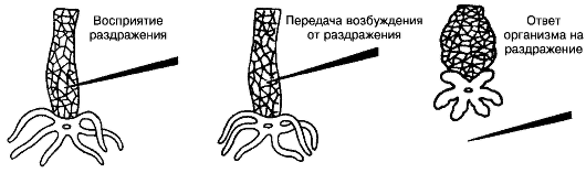
Irritability, reflexes. Hydra is able to sense touch, temperature changes, the appearance of various dissolved substances in water and other irritations. This causes her nerve cells to become excited. If you touch the hydra with a thin needle, then the excitement from irritation of one of the nerve cells is transmitted along the processes to other nerve cells, and from them to the skin-muscle cells. This causes muscle fibers to contract, and the hydra shrinks into a ball. 14
In this example, we get acquainted with a complex phenomenon in the animal’s body - a reflex. Reflex consists of three successive stages: perception of irritation, transmission of excitation from this irritation through nerve cells and the body’s response with some action. Due to the simplicity of the hydra's organization, its reflexes are very uniform. In the future we will become familiar with much more complex reflexes in more highly organized animals.
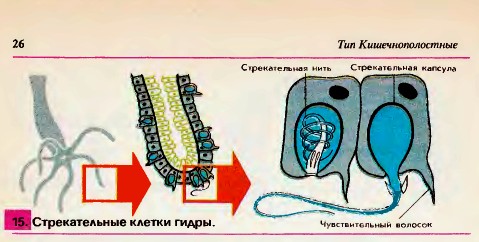
Stinging cells. The entire body of the hydra and especially its tentacles are seated with a large number of stinging, or nettle, cells 15. Each of these cells has a complex structure. In addition to cytoplasm and kernels it contains a bubble-like stinging capsule, inside which a thin tube is coiled - the stinging thread. A sensitive hair sticks out from the cell. As soon as a crustacean, small fish or other small animal touches a sensitive hair, the stinging thread quickly straightens, its end is thrown out and pierces the victim. Through a channel passing inside the thread, poison enters the body of the prey from the stinging capsule, causing the death of small animals. As luck would have it, a lot of stinging cells shoot out at once. then the hydra uses its tentacles to pull the prey to its mouth and swallows it. The stinging cells also serve the hydra for protection. Fish and aquatic insects do not eat hydras, which burn enemies. The poison from the capsules is similar in its effect on the body of large animals to the poison of weed.
1. What environmental factors does the life of a hydra depend on?
2. How is hydra adapted to its habitat?
3. name the cells of the outer layer of hydra. What significance do they have in her life?
4. Why do many fish, having captured a hydra in their mouths, throw it away?
5. What is a reflex?
6. using fig. 14
, tell us how the hydra reflex appears. What is the significance of hydra's reflexes?
Biology: Animals: Textbook. for 7th grade avg. school / B. E. Bykhovsky, E. V. Kozlova, A. S. Monchadsky and others; Under. ed. M. A. Kozlova. - 23rd ed. - M.: Education, 2003. - 256 p.: ill.
Calendar and thematic planning in biology, video in biology online, Biology at school download
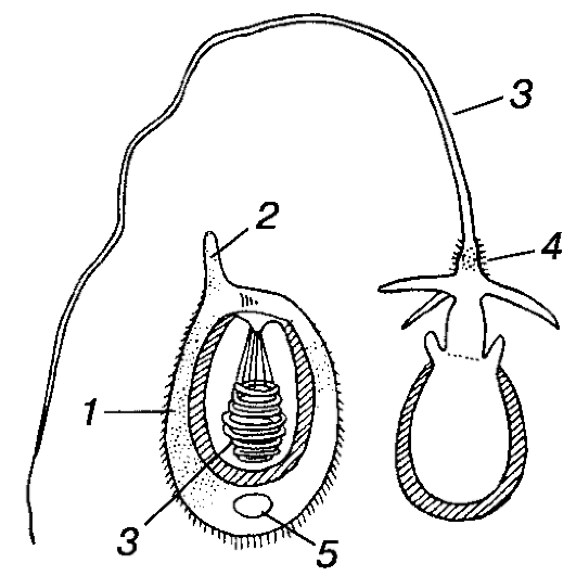
Lesson content lesson notes supporting frame lesson presentation acceleration methods interactive technologies Practice tasks and exercises self-test workshops, trainings, cases, quests homework discussion questions rhetorical questions from students Illustrations audio, video clips and multimedia photographs, pictures, graphics, tables, diagrams, humor, anecdotes, jokes, comics, parables, sayings, crosswords, quotes Add-ons abstracts articles tricks for the curious cribs textbooks basic and additional dictionary of terms other Improving textbooks and lessonscorrecting errors in the textbook updating a fragment in a textbook, elements of innovation in the lesson, replacing outdated knowledge with new ones Only for teachers perfect lessons calendar plan for the year guidelines discussion programs Integrated LessonsA characteristic feature of coelenterates is the presence of stinging cells in the integument (Fig. 93). They develop from intermediate cells and contain a special oval stinging capsule with dense walls. The capsule is filled with liquid, and at one end of the capsule its wall is pushed inward in the form of a very thin but hollow process, which twists into the capsule into a spirally curled stinging thread. The stinging cells serve the hydra as a weapon of attack and defense.
On the outer surface of the cell there is a thin sensitive hair - cnidocil. Studying stinging cells using an electron microscope showed significant complexity in the structure of the cnidocil (Fig. 93). It consists of a long flagellum surrounded by 18-22 thin finger-like outgrowths of the cytoplasm - microvilli. In structure, the cnidocil flagellum is very similar to the flagella and cilia of protozoa, but unlike them, it is motionless. When prey or an enemy touches the flagellum, the latter deflects and touches one or more microvilli, which leads to the excitation of the stinging cell. At the same time, the stinging capsule throws out an elastic thread that turns outward and straightens out like an arrow. The thread, like a harpoon, is seated with back-facing spines, and at the base it bears larger spines. Thread pricks are poisonous and can paralyze small animals. After the thread is thrown out, the stinging cell dies.
Hydra has several categories of capsules, the functions of which are different. The large capsules considered, which serve to pierce the covers and damage prey, are called penetrants (Fig. 93). Much smaller ones - volvents - have short spirally twisted threads that wrap around various protrusions (setae, hairs, etc.) on the body of the prey and in this way hold it. Finally, elongated stinging capsules - glutinants - are glued to the body of the prey with long sticky threads.