The vital activity of our body is ensured by the coordinated functioning of our organ systems.
An important role in the regulation and performance of all functions is played by the human excretory organs.
Nature has awarded us with special organs that help remove metabolic products from the body.
What excretory organs does a person have?
The human organ system consists of:
- kidney,
- Bladder,
- ureters,
- urethra.
In this article we will take a detailed look at the human excretory organs and their structure and functions.
Kidneys
These paired organs are located on the back wall of the abdominal cavity, on both sides of the spine. The kidney is a paired organ.
Outwardly she has bean-shaped and inside - parenchymal structure. Length one kidney no more than 12 cm, and width- from 5 to 6 cm. Normal weight kidneys do not exceed 150-200 g.
Structure
The membrane that covers the outside of the kidney is called fibrous capsule. On a sagittal section, two different layers of substance can be seen. The one located closer to the surface is called cortical, and the substance occupying the central position is cerebral.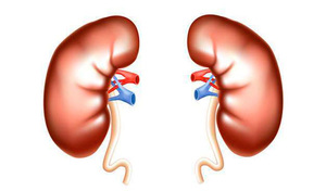
They have not only external differences, but also functional ones. On the side of the concave part there are hilum of the kidney and pelvis, and ureter.
Through the renal hilum, the kidney communicates with the rest of the body through the incoming renal artery and nerves, as well as the outgoing lymphatic vessels, the renal vein and the ureter.
The collection of these vessels is called renal pedicle. Inside the kidney there are renal lobes. There are 5 pieces in each kidney. The renal lobes are separated from each other by blood vessels.
In order to clearly understand the functions of the kidneys, it is necessary to know them. microscopic structure.
The main structural and functional unit of the kidneys is nephron.
Number of nephrons in the kidney reaches 1 million. The nephron consists of renal corpuscle, which is located in the cortex, and tubule systems, which ultimately flow into the collecting duct.
In the nephron there are also 3 segments:
- proximal,
- intermediate,
- distal.
Segments along with the ascending and descending limbs of the loop of Henle lie in the renal medulla.
Functions
Along with the main excretory function, the kidneys also provide and perform:
- maintaining a stable level blood pH, its circulating volume in the body and the composition of the intercellular fluid;
- thanks to metabolic function, human kidneys carry out synthesis of many substances, important for the life of the body;
- blood formation, by producing erythrogenin;
- synthesis of such hormones, such as renin, erythropoietin, prostaglandin.
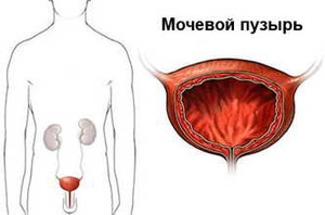
Bladder
The organ that stores urine that passes through the ureters and removes it through the urethra is called bladder . This is a hollow organ that is located in the lower abdomen, just behind the pubis.
Structure
The bladder is round in shape, in which there are
- top,
- body,
- neck
 The latter narrows, thus passing into the urethra. When filled, the walls of the organ stretch, signaling the need to empty.
When the bladder is empty, its walls thicken, and the mucous membrane gathers into folds. But there is a place that remains unwrinkled - this is the triangular area between the opening of the ureter and the opening of the urethra.
Functions
The bladder performs the functions:
- temporary accumulation of urine;
- urine excretion— the volume of urine accumulated in the bladder is 200-400 ml. Every 30 seconds, urine flows into the bladder, but the time of entry depends on the amount of liquid drunk, temperature, and so on;
- thanks to mechanoreceptors that are located in the wall of the organ, it is carried out control of the amount of urine in the bladder. Their irritation serves as a signal to contract the bladder and remove urine out.
Ureters
The ureters are thin ducts that connects the kidney and bladder. Their length is no more than 30 cm, and diameter from 4 to 7 mm.
Structure
The tube wall has 3 layers:
For the prevention of diseases and treatment of the kidneys and urinary system, our readers recommend Father George's Monastic Tea. It consists of 16 of the most beneficial medicinal herbs that have extremely high efficiency in cleansing the kidneys, in the treatment of kidney diseases, urinary tract diseases, as well as in cleansing the body as a whole. Doctors' opinion..."
- external (from connective tissue),
- muscular and internal (mucous membrane).
One part of the ureter is located in the abdominal cavity, and the other in the pelvic cavity. If there are difficulties in the outflow of urine (stones), the ureter may expand in some area up to 8 cm.
Functions
The main function of the ureter is urine outflow accumulated in the bladder. Due to contractions of the muscle membrane, urine moves through the ureter into the bladder.
Urethra
In women and men, the urethra differs in its structure. This is due to the difference in genital organs.
Structure
The canal itself consists of 3 membranes, like the ureter. Because women have a urethra 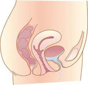 In short, than in men, women are more often exposed to various diseases and inflammations of the urogenital tract.
Functions
- In men the channel performs several functions: excretion of urine and sperm. The fact is that the vas deferens ends in the canal tube, through which sperm flows through the canal into the head of the penis.
- Among women The urethra is a tube 4 cm long and performs only the function of excreting urine.
How are primary and secondary urine formed?
The process of urine formation includes three interconnected stages:
- glomerular filtration,
- tubular reabsorption,
- tubular secretion.
First stage - glomerular filtration is the process of transition of the liquid part of the plasma from the capillaries of the glomerulus into the lumen of the capsule. In the lumen of the capsule there is a filtration barrier, which contains pores in its structure that selectively allow dissimilation products and amino acids to pass through, and also prevent the passage of most proteins.
During glomerular filtration, it is formed ultrafiltrate, representing primary urine. It is similar to blood plasma, but contains few proteins.
During the day, a person produces from 150 to 170 liters of primary urine, but only 1.5-2 liters turns into secondary urine, which is excreted from the body.
The remaining 99% returns to the blood.
Mechanism formation of secondary urine consists in the passage of ultrafiltrate through segments  nephron and renal tubules. The walls of the tubules consist of epithelial cells, which gradually absorb back not only a large number of water, but also all the substances necessary for the body.
The reabsorption of proteins is explained by their large size. All substances that are toxic and harmful to our body remain in the tubules and are then excreted in the urine. This final urine is called secondary. This whole process is called tubular reabsorption.
Tubular secretion is a set of processes due to which substances to be excreted from the body are secreted into the lumen of the nephron tubules. That is, this secretion is nothing more than a reserve process of urine formation.
Selection- part of the metabolism carried out by removing from the body the final and intermediate products of metabolism, foreign and excess substances to ensure the optimal composition of the internal environment and normal life activity.
excretory system organs
| organ | secreted substance |
| kidneys | excess water inorganic and organic substances end products of metabolism |
| lungs | water vapor some volatile substances (for example, ether and chloroform vapors during anesthesia, alcohol vapors during intoxication) |
| salivary glands | heavy metals drugs (for example, morphine and quinine) alien organic compounds |
| liver | products of nitrogen metabolism (urea) hormones (for example, thyroxine) hemoglobin breakdown products medications |
| pancreas | heavy metals medicinal substances |
| intestinal glands | heavy metals medicinal substances |
| leather | lactic acid urea uric acid |
Excretion products
In the process of life, end products of metabolism are formed in the body. Most of them are non-toxic to the body (for example, carbon dioxide and water).
However, during the oxidation of proteins and other nitrogen-containing products, ammonia is formed - one of the end products of nitrogen metabolism. It is toxic to the body, so it is quickly eliminated from the body. Dissolving in water, ammonia turns into a low-toxic compound -- urea.
Urea is formed mainly in the liver. The amount of urea excreted in the urine per day is approximately 50 - 60 g. Thus, the products of nitrogen metabolism are practically excreted in the urine in the form of urea.
Part of the nitrogen is excreted from the body in the form uric acid, creatine and creatinine. These substances are the main nitrogen-containing components of urine.
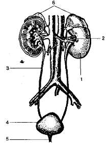
urinary system
Human urinary system- a system of organs that form, accumulate and secrete urine.
STRUCTURE OF THE URINARY SYSTEM:
- two kidneys
- two ureters
- bladder
- urethra
Rice. Organs of the urinary system
kidney function
The role of the kidneys in the body is not limited to the release of end products of nitrogen metabolism and excess water. The kidneys are actively involved in maintaining homeostasis in the body.
- osmoregulation- maintaining osmotic pressure in the blood and other body fluids;
- ion regulation- regulation of the ionic composition of the internal environment of the body;
- maintaining the acid-base balance of blood plasma (pH = 7.4);
- regulation of blood pressure;
- endocrine function: synthesis and release into the blood biologically active substances:
- renina regulating blood pressure;
-erythropoietin, regulating the rate of red blood cell formation; - participation in metabolism;
- excretory function: excretion from the body of the final products of nitrogen metabolism, foreign substances, excess organic matter(glucose, amino acids, etc.).
Kidney structure
Kidneys- bean-shaped parenchymal organs located on the dorsal side on the sides of the lumbar spine.
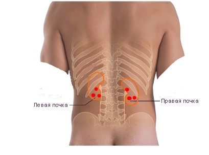
Rice. Kidney location
Each bud measures approximately 4 x 6 x 12 cm and weighs approximately 150 g.
The kidney is surrounded by three membranes (capsules):
- fibrous capsule- internal thin and dense shell;
in the inner part of this capsule there are smooth muscle cells, due to their slight contraction, the pressure necessary for filtration processes is maintained in the kidney. - fat capsule - middle shell;
fatty tissue is more developed on the back side of the kidney. Function: elastic fixation of the kidney in the lumbar region; thermoregulation; mechanical protection (shock absorption). When you lose weight and reduce the volume of fatty tissue, mobility or prolapse of the kidneys may occur. - renal fascia- the outer membrane covering the kidney with a fat capsule and adrenal glands. The fascia holds the kidney in a certain position. Connective tissue fibers pass from the fascia to the fibrous capsule through the fatty tissue.
The kidney parenchyma includes:
- cortical layer (outer layer) 5 - 7 mm thick;
- medulla (inner layer);
- renal pelvis.

Rice. Kidney anatomy
The cortex is located on the periphery of the kidney and in the form of pillars ( Bertini speakers) penetrates deeply into the medulla. The medulla is divided into 15 - 20 by renal columns renal pyramids, with their tops facing inward and their bases facing outward. The pyramid of the medulla together with the adjacent cortical substance form kidney lobe.

Rice. Structure of the kidney and nephron
Pelvis- the central hollow part of the kidney into which secondary urine from all nephrons drains. The wall of the pelvis consists of mucous, smooth muscle and connective tissue membranes.
The ureter originates from the renal pelvis, carrying the resulting urine to the bladder.
Ureters
Ureters- hollow tubes connecting the kidneys to the bladder.
Their wall consists of epithelial, smooth muscle and connective tissue layers.
Contraction of smooth muscles causes urine to flow from the kidneys to the bladder.
bladder
Bladder- a hollow organ capable of strong stretching.
Rice. Bladder
Bladder function:
- urine accumulation;
- control of the amount of urine in the bladder;
- excretion of urine.
Like all hollow organs, the bladder has a three-layer wall:
- inner layer of transitional epithelium;
- medium thick smooth muscle layer;
- outer connective tissue layer.
urethra
Urethra- a tube connecting the bladder to the external environment.
The canal wall consists of 3 membranes: epithelial, muscular and connective tissue.
The outlet of the urethra is called urethra.
Two sphincters block the lumen of the canal at the junction with the bladder and in the urethra.
In women, the urethra is short (about 4 cm), and it is easier for infections to enter the female genitourinary system.
In men, the urethra serves to secrete not only urine, but also sperm.
nephron structure
The structural and functional unit of the kidney is nephron.
Each human kidney contains about 1 million nephrons.
The main processes that determine the various functions of the kidneys occur in the nephron.
Structural parts of the nephron:
- renal (Malpighian) corpuscle:
- capillary (renal) glomerulus (+ afferent and efferent arteries)
- Bowman-Shumlyansky capsule (= nephron capsule): formed by two layers of epithelial cells; the lumen of the capsule becomes a convoluted tubule;
- convoluted tubule of the first order (proximal): its walls have a brush border - a large number of microvilli facing the lumen of the tubule.
- loop of Henle: descends into the medulla, and then turns 180 degrees and returns to the cortex;
- convoluted tubule of the second order (distal): the walls of the loop of Henle and the distal convoluted tubule are without villi, but have strong folding;
- collecting tube.
In different parts of the nephron, different processes occur that determine the functions of the kidneys. The location of the parts of the nephron is also connected with this:
- the glomerulus, capsule and convoluted tubules are located in the cortex;
- The loop of Henle and collecting ducts are located in the medulla.
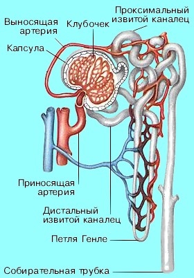
Rice. Nephron vessels
Beginning in the renal cortex, the collecting ducts pass through the medulla and open into the cavity of the renal pelvis.
Circulatory system of the kidneys
Blood reaches the kidneys through the renal arteries (branches of the abdominal aorta). The arteries branch strongly and form a vascular network. An afferent arteriole enters each renal capsule, there it forms a capillary network - the renal glomerulus - and leaves the capsule in the form of a thinner efferent arteriole. This creates high blood pressure in the capillaries of the glomerulus to filter the liquid part of the blood and form primary urine. The pressure in the capillaries of the glomerulus is quite stable, its value remains constant even with an increase general level pressure. Consequently, the filtration rate also remains virtually unchanged.
After leaving the glomerulus, the efferent arteriole again breaks up into capillaries, forming a dense network around the convoluted lot. Thus, most of Blood in the kidney passes through the capillaries twice - first in the glomerulus, then at the tubules.
Blood is carried out of the kidneys through the renal veins, which flow into the inferior vena cava.
PROCESSES OCCURRING IN THE KIDNEYS
- ultrafiltration of fluid in the renal glomeruli;
- reabsorption (reabsorption);
- urine excretion.
ULTRAFILTRATION OF FLUID IN RENAL glomeruli
The initial stage of urine formation occurs in the glomeruli - ultrafiltration from the blood plasma into the capsule of the renal glomerulus of all low-molecular components of the blood plasma.
Moreover, in the process tubular secretion nephron epithelial cells capture certain substances from the blood and intercellular fluid and transfer them into the lumen of the tubule.
Thus, approximately 170 liters are formed per day primary urine.
The composition of primary urine is similar to that of blood plasma, devoid of protein:
- mineral salts
- low molecular weight compounds (including toxins, amino acids, glucose, vitamins)
- NO PROTEIN (trace)
- NO BLOOD ELEMENTS
REABSORPTION (REVER SUCTION)
The second stage is associated with the reabsorption into the blood capillaries of all substances valuable to the body: water, ions ( N a+ Na+, C l− Cl−, H C O− 3 HCO3−), amino acids, glucose, vitamins, proteins, microelements. Reabsorption of sodium and chlorine is the most significant process in terms of volume and energy consumption.
Reabsorption occurs during the passage of primary urine through the convoluted tubule system. For this purpose, the efferent arteriole breaks up again into a network of capillaries that entangle the tubules: through their thin walls, the substances needed by the body are reabsorbed.
A small amount of protein filtered in the glomeruli is reabsorbed by the cells of the proximal tubules. The excretion of proteins in the urine is normally no more than 20 - 75 mg per day, and in case of kidney disease it can increase to 50 g per day. An increase in the excretion of proteins in the urine (proteinuria) may be due to a violation of their reabsorption or an increase in filtration.
As a result of filtration, reabsorption and secretion, only 1.5 liters remain from 180 liters of primary urine concentrated solution"unnecessary" substances - secondary urine.
Composition of secondary urine:
- toxins
- metabolic products (including drug residues)
EXCRETION OF SUBSTANCES
Secondary urine enters the renal pelvis through the collecting ducts.
The average person produces approximately 1.5 liters of urine per day.
From the kidneys, urine travels through the ureters to the bladder.
The capacity of the bladder is on average 600 ml.
Usually the contents of the bladder are sterile.
The wall of the bladder has a muscle layer that, when contracted, causes urination.
Urination- a voluntary (consciously controlled) reflex act triggered by tension receptors in the wall of the bladder, sending a signal to the brain about the filling of the bladder.
The flow of urine as it is released from the bladder is regulated by the circular sphincter muscles. When the bladder begins to empty, the bladder sphincter relaxes and the bladder wall muscles contract, creating a flow of urine.
During the metabolism of proteins and nucleic acids Various products of nitrogen metabolism are formed: urea, uric acid, creatinine, etc.
When the excretion of uric acid is impaired, gout develops.
Endocrine kidney function
The kidneys produce:
- ammonia: excreted in urine;
- renin, prostaglandins, glucose synthesized in the kidney: enter the blood.
Ammonia enters primarily into the urine. Some of it penetrates into the blood, and there is more ammonia in the renal vein than in the renal artery.
regulation of kidney function
- Vasopressin (= antidiuretic hormone (ADH) - a hormone of the hypothalamus that accumulates in the neurohypophysis):
increases the reabsorption of water by the kidney, thereby increasing the concentration of urine and decreasing its volume - Aldosterone (hormone of the adrenal cortex):
increased reabsorption N a+ Na+atWithAndlenAndeWitheToRetsAndAndincreased secretion K^+$
- Natriuretic hormone (atrial hormone):
increased secretion N a+ Na+
decreased potassium excretion.
Thematic assignments
A1. Decomposition products of similar composition are removed through
1) skin and lungs
2) lungs and kidneys
3) kidneys and skin
4) digestive tract and kidneys
A2. The organs of the excretory system are located
1) in the chest cavity
3) outside body cavities
2) in the abdominal cavity
4) in the pelvic cavity
A3. The integral structural unit of the kidney is
1) neuron
2) nephron
3) capsule
4) convolute
A4. If the process of excretion of decay products is disrupted, the following accumulates in the body:
1) salts of sulfuric acid
3) glycogen
2) excess proteins
4) urea or ammonia
A5. Function of the capillary (malpighian) glomerulus:
1) blood filtration
3) water absorption
2) urine filtration
4) lymph filtration
A6. Conscious retention of urination is associated with the following activities:
1) medulla oblongata
3) spinal cord
2) midbrain
4) cerebral cortex
A7. Secondary urine differs from primary urine in that secondary urine does not contain:
1) glucose
2) urea
3) salts
4) K ions + and Ca 2 +
A8. Primary urine is formed from:
1) lymph
2) blood
3) blood plasma
4) tissue fluid
A9. Presence in urine may be a symptom of kidney disease.
1) sugar
2) potassium salts
3) sodium salts
4) urea
A10. Humoral regulation of kidney activity is carried out with the help of
1) enzymes
2) vitamins
3) amino acids
4) hormones
IN 1. Select symptoms that may indicate kidney disease
1) the presence of proteins in the urine
2) the presence of uric acid in the urine
3) increased glucose content in secondary urine
4) low leukocyte count
5) increased content of leukocytes
6) increased daily amount of urine excreted
AT 2. Which of the following applies to the nephron?
1) renal pelvis
2) ureter
3) capillary glomerulus
4) capsule
5) bladder
6) convoluted lot
During the life of the body, the breakdown of proteins, fats and carbohydrates occurs in the tissues with the release of energy. The final decomposition products are water, carbon dioxide, ammonia, urea, uric acid, phosphate salts and other compounds. From the tissues, these dissimilation products pass into the blood, are carried by the blood to the excretory organs and through them are removed from the body. The removal of these substances involves the lungs, skin, digestive system and organs of the urinary system.
Most of the breakdown products are excreted through the urinary system. This system includes the kidneys, ureters, bladder and urethra.
Thanks to their activity in the human body, the kidneys are involved in maintaining a constant volume of body fluids, their osmotic pressure and ionic composition; regulation of acid-base balance; release of nitrogen metabolism products and foreign substances; saving or excretion of various organic substances (glucose, amino acids, etc.) depending on the composition of the internal environment; metabolism of carbohydrates and proteins; secretion of biologically active substances (renin hormone); hematopoiesis. The kidneys have a wide range of functional adaptation to the body's needs in maintaining homeostasis, since they are capable of significantly varying the qualitative composition of urine, its volume, osmotic pressure and pH.
The right and left kidneys, each about 150 g, are located in the retroperitoneal space on the sides of the spinal column at the level of the lumbar vertebrae. The outside of the buds is covered with a dense membrane. On the inner concave side there is a “gate” of the kidney through which urine passes. rosary, renal arteries and veins, lymphatic vessels and nerves. A cross-section of the kidney shows that it consists of two layers: the outer layer (the darker one is the cortex), the inner layer is the medulla.
The kidney has a complex structure and consists of approximately 1 million structural and functional units - nephrons, the space between which is filled with connective tissue.
Nephrons are complex microscopic formations that begin with a double-walled glomerular capsule (Shumlyansky-Bowman capsule), inside which there is a renal corpuscle (Malpighian corpuscle). Between the layers of the capsule there is a cavity that passes into the convoluted (primary) urinary tubule. It reaches the border of the cortex and medulla of the kidney. At the border, the tubule narrows and straightens. In the medulla of the kidney it forms a loop and returns to the cortex of the kidney... Here it again becomes convoluted (secondary) and opens into the collecting duct. The collecting ducts, merging, form common excretory ducts, which pass through the medulla of the kidney to the tips of the papillae, protruding into the cavity of the pelvis. The pelvis passes into the ureter.
 How is urine formed in nephrons? In a simplified form, this happens as follows. When blood passes through the capillaries of the glomeruli, water and substances dissolved in it are filtered from its plasma through the capillary wall into the capsule cavity, with the exception of large molecular compounds and blood elements. Consequently, large proteins do not enter the filtrate. molecular weight. But such metabolic products as urea, uric acid, ions come here inorganic substances, glucose and amino acids. This filtered liquid is called primary urine. Filtration is carried out due to high pressure in the capillaries of the glomeruli - 60-70 mm Hg. Art., which is two or more times higher than in the capillaries of other tissues. It is created due to the different sizes of the lumens of the afferent (wide) and efferent (narrow) vessels.
During the day, a huge amount of primary urine is formed - 150-180 liters. Such intensive filtration is possible due to the large amount of blood that flows through the kidneys during the day - 1500-1800 liters; large surface area of the walls of the capillaries of the glomeruli - 1.5 m 2; high blood pressure in them, which creates a filtering force, and other factors.
From the glomerular capsule, primary urine enters the primary tubule, which is densely entwined with secondarily branched blood capillaries. In this part of the tubule, absorption (reabsorption) of most of the water and a number of substances into the blood occurs; glucose, amino acids, low molecular weight proteins, vitamins, sodium, potassium, calcium, chlorine ions. That part of the primary urine that remains at the end of its passage through the tubules is called secondary. It contains; urea, uric acid, ammonia, sulfates, phosphates, sodium, potassium, etc. Consequently, in secondary urine, with normal kidney function, there are no proteins and sugars. Their appearance there indicates a violation of the kidneys, although with excessive consumption of simple carbohydrates (over 100 g per day), sugars may appear in the urine even with healthy kidneys.
A little secondary urine is formed - about 1.5 liters per day. The rest of the primary urine liquid from a total amount of 150-180 liters is absorbed into the blood through the cells of the walls of the urinary tubules. Total surface their size is 40-50 m2.
The kidneys do a lot of work non-stop. Therefore, with a relatively small size, they consume a lot of oxygen and nutrients, which indicates large energy expenditures during the formation of urine. Thus, they consume 8-10% of all oxygen absorbed by a person at rest. More energy is spent per unit mass in the kidneys than in any other organ.
Urine collects in the bladder. As it accumulates, its walls stretch. This is accompanied by irritation of the nerve endings located in the walls of the bladder. Signals are sent to the central nervous system and the person feels the urge to urinate. It is carried out through the urethra and is under the control of the nervous system.
An efficient excretion process as an integral part of metabolism - important condition maintaining homeostasis. It ensures the liberation of the body from unnecessary and harmful metabolic products, as well as from foreign and toxic substances (alcohol, drugs, medicines, etc.) that came from outside. Organs of excretion are kidneys, lungs, leather(her sweat glands), salivary glands, gastrointestinal tract, liver (Fig. 96).
Lungs remove carbon dioxide, water vapor, and some volatile substances (air after anesthesia, alcohol) from the body. The volume of water vapor that is excreted by the lungs depends on temperature and humidity. environment, level of physical and emotional stress.
Salivary glands remove medicinal substances (for example, aspirin), various foreign organic compounds that have entered the body.
Performs an important excretory function liver. Harmful substances that accidentally enter the intestines with food, as well as rotting products, are absorbed from it into the blood and enter the liver. In it they are neutralized, lose their toxicity and are excreted as part of bile through intestines, which also frees the body from unnecessary and harmful metabolic products, undigested food debris and pathogens.
Sweat glands remove excess water, mineral salts, heavy metal salts that can enter the body with food, some products of protein metabolism (for example, urea), and during heavy physical work - lactic acid.
Kidneys- the main excretory organ in the human body. They perform two main functions: removing toxic products of protein metabolism and nucleic acid metabolism from the body; maintain the normal water-salt composition of body fluids (blood, lymph, intercellular fluid). In addition, toxic and foreign substances are eliminated through the kidneys. Material from the site
During the breakdown of nucleic acids and amino acids in cells, ammonia is formed, which is a poison for human body. Getting into the liver with the bloodstream, it turns into a less toxic substance - urea, which is eliminated through the kidneys aqueous solution. This process is closely related to the exchange of water and salts in the body. During the day, approximately 2 liters of water with urea, sodium chloride and a small amount of other substances dissolved in it are removed from the human body through urine.
All physiological systems that perform the excretory function constantly interact with each other. For example, when, to maintain optimal body temperature during hot weather or heavy physical activity, a large amount of water evaporates from the surface of the skin through sweat, the excretion of the latter in urine is significantly reduced. On the contrary, at low temperatures, when the evaporation of water through the skin is reduced, the kidneys excrete more water.