These tissues are excitable tissues, i.e. they are capable of responding to stimulation with stimulation and conducting it at a distance.
Muscle tissue
By the origin and structure of muscle tissue are significantly different from each other, but they are united by the ability to contract, which provides the motor function of organs and the body as a whole. The muscle elements are elongated in length and are associated either with other muscle elements or with supporting formations.
Distinguish between smooth, striated muscle tissue and muscle tissue of the heart (Fig. 5).
Smooth muscle tissue.
This tissue is formed from mesenchyme. The structural unit of this tissue is a smooth muscle cell. It has an elongated fusiform shape and is covered with a cell membrane. These cells are tightly adjacent to each other, forming layers and groups separated by a loose unformed connective tissue.
The cell nucleus has an elongated shape and is located in the center. Myofibrils are located in the cytoplasm, they go along the periphery of the cell along its axis. They consist of thin threads and are a contractile element of the muscle.
Cells are located in the walls of blood vessels and most of the internal hollow organs (stomach, intestines, uterus, bladder). Smooth muscle activity is regulated by the autonomic nervous system. Muscle contractions do not obey the will of a person and therefore smooth muscle tissue is called involuntary musculature.
Striated muscle tissue.
This tissue was formed from myotomas, derivatives of the mesoderm. The structural unit of this tissue is the striated muscle fiber. This cylindrical body is a symplast. It is covered with a shell - sarcolem, and the cytoplasm is called - sarcoplasma, in which there are numerous nuclei and myofibrils. Myofibrils form a bundle of continuous fibers extending from one end of the fiber to the other parallel to its axis. Each myofibril consists of disks having different chemical composition and under the microscope seeming dark and light. The homogeneous discs of all myofibrils coincide, and therefore the muscle fiber appears to be striated. Myofibrils are the contractile apparatus of muscle fiber.
The whole skeletal muscles are built from the striated muscle tissue. The muscles are arbitrary, because its contraction can occur under the influence of neurons of the motor zone of the cerebral cortex.
Muscular tissue of the heart.
Myocardium - the middle layer of the heart - is built from striated muscle cells (cardiomyocytes). There are two types of cells: typical contractile cells and atypical cardiac myocytes that make up the conduction system of the heart.
Typical muscle cells perform a contractile function; they are rectangular in shape, 1-2 nuclei are in the center, myofibrils are located on the periphery. Between adjacent myocytes there are insertion discs. With their help, myocytes collect in muscle fibers, separated by a thin-fibrous connective tissue. Between adjacent muscle fibers are connecting fibers that provide contraction of the myocardium as a whole.
The conduction system of the heart is formed by muscle fibers consisting of atypical muscle cells. They are larger than contractile, richer in sarcoplasm, but poorer than myofibrils, which often intersect. The kernels are larger and not always in the center. The fibers of the conducting system are surrounded by a dense plexus of nerve fibers.
Nerve tissue.
Nerve tissue consists of nerve cells with a specific function, and neuroglia, which performs protective, trophic and supporting functions. Comes from the ectoderm.
A nerve cell, or neuron, is characterized by the ability to perceive irritations, come into a state of excitement and transmit it to other cells of the body. Due to this, the interconnection of organs and tissues, the regulation of all body functions and its adaptation to the environment.
Nerve cells have a different shape and size and consist of a body and processes (Fig. 6).
The processes of the nerve cell are divided into two types:
· Neurites, or axons along which excitation (impulse) is transmitted from the cell body to the periphery. The axon always leaves one cell and ends with the end device in the working organ or on another neuron.
· Dendrites - processes along which an impulse is transmitted from the periphery to the cell body. There are many of them and they branch.
By the number of processes, nerve cells are divided into three types (Fig. 7):
· Unipolar - cells with one process. Not found in humans.
· Bipolar - have one neuritis in the central nervous system and one dendrite going to the periphery. They are located in the spinal nerve nodes.
· Multipolar - have one neuritis and many dendrites. A person has them the most.
The nucleus of the nerve cell has a rounded shape and is located in the center.
In the cytoplasm of neurons there are neurofibrils, which are thin filaments. In the body of the nerve cell they form a dense network. In the processes of neurofibrils are parallel to each other.
Neuroglia represented by cells of various shapes with a large number of processes. These cells are more than nerve cells.
Nerve fibers. The processes of nerve cells with membranes are called nerve fibers. Distinguish between myelin (pulp) and bezmyelinovye (serene). The processes are located in the center of the nerve fiber and are called the axial cylinder, which is covered by a membrane formed by neuroglia cells (lemmocytes).
Myelin-free the fibers are an axial cylinder, covered only with a membrane of lemmocytes.
Myelin - much thicker. They also consist of an axial cylinder, but have two layers of the shell: the inner, the thicker - myelin, and the outer, thin, consisting of lemmocytes. Outside, myelin fiber is covered with a thin connective tissue sheath - unacceptable.
Nerve endings. All nerve fibers end with nerve endings. There are three groups:
· Efferent. They can be of two types: motor and secretory. The motor endings are the terminal apparatuses of the axons of the somatic and autonomic nervous system.
· Sensitive (receptors) are the terminal devices of dendrites of sensitive neurons. They are divided into free ones, consisting of a branching of the axial cylinder, and not free, containing all the components of the nerve fiber covered by the capsule.
· End processes, forming interneuron synapses that connect neurons to each other.
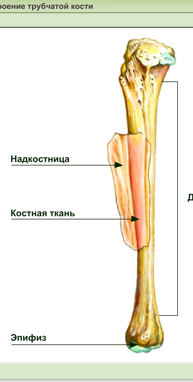
Bone - a kind of connective tissue from which bones are built - the organs that make up the skeleton of the human body. Bone tissue is important in terms of the musculoskeletal system and other body systems.
Bone tissue consists of interacting structures:
Bone cell
Intercellular Organic Bone Matrix (Organic Bone Skeleton),
The main mineralized intercellular substance.
Cells occupy only 1-5% of the total bone tissue of an adult human skeleton. There are four types of bone cells.
Osteoblasts - germ cells that perform the function of creating bone. They are located in areas of bone formation on the external and internal surfaces of the bone.
Osteoclasts - cells that perform the function of resorption, bone destruction. The joint function of osteoblasts and osteoclasts is the basis of a continuous controlled process of bone destruction and reconstruction. This process of bone remodeling underlies the body's adaptation to diverse physical exertion by choosing the best combinations of stiffness, elasticity and elasticity of bones and skeleton.
Osteocytes - cells originating from osteoblasts. They are completely walled up in the intercellular substance and contact the processes with each other. Osteocytes provide metabolism (of proteins, carbohydrates, fats, water, minerals) of bone tissue. Undifferentiated mesenchymal bone cells (osteogenic cells, contour cells). They are located mainly on the outer surface of the bone (at the periosteum) and on the surfaces of the internal spaces of the bone. Of these, new osteoblasts and osteoclasts are formed.
Intercellular substance it is represented by an organic intercellular matrix built of collagen (ossein) fibers (≈90-95%) and the main mineralized substance (≈5-10%).
The collagen of the extracellular matrix of bone tissue differs from the collagen of other tissues in the high content of specific polypeptides. Collagen fibers are generally parallel to the direction of the level of the most likely mechanical stresses on the bone and provide bone resilience and elasticity.
Main substance consists mainly of extracellular fluid, glycoproteins and proteoglycans (chondroitin sulfates, hyaluronic acid). The function of these substances is not yet clear, but it is certain that they are involved in controlling the mineralization of the main substance — the movement of the mineral components of the bone.
Mineralsplaced in the main substance in the organic matrix of the bone are crystals built mainly of calcium and phosphorus. The ratio of calcium / phosphorus is normally ≈1.3-2.0. In addition, magnesium, sodium, potassium, sulfate, carbonate, hydroxyl and other ions that can take part in the formation of crystals were found in the bone. Each collagen fiber of a compact bone is constructed of periodically repeating segments. The fiber segment length is ≈64 nm (64 10-10 m). Hydroxyapatite crystals are adjacent to each fiber segment, tightly encircling it.
 In addition, segments of adjacent collagen fibers overlap. Accordingly, like bricks when laying walls, hydroxyapatite crystals overlap each other. Such a close fit of collagen fibers and hydroxyapatite crystals, as well as their overlap, prevents the "destruction of the shear" of the bone under mechanical stress. Collagen fibers provide elasticity, elasticity of the bone, its resistance to stretching, while crystals provide its strength, rigidity, its resistance to compression. Bone mineralization is associated with features of bone tissue glycoproteins and osteoblast activity.
Distinguish coarse fiber and lamellar bone tissue .
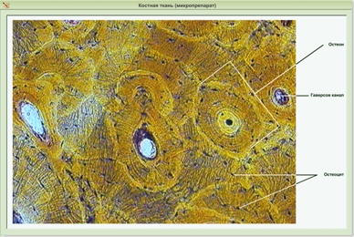
In coarse-fibrous bone tissue (prevails in embryos; in adult organisms, it is observed only in the area of \u200b\u200bcranial sutures and tendon attachment sites), the fibers go disordered. In lamellar bone tissue (bones of adult organisms), fibers grouped into separate plates are strictly oriented and form structural units called osteons.
Osteon is a three-dimensional cylindrical system of concentrically located bone plates and osteocytes surrounding the central channel of the osteon. In bone plates, ossein fibrils are closely and parallel to each other. Bone-plate cylinders are inserted one into the other. In adjacent concentric bone plates, axial new fibrils go at a different angle. Due to this, the exceptional strength of osteons is achieved. The complex construction of osteons is formed in the process of histogenesis of bone tissue and its constant restructuring. Some osteons are destroyed. The remnants of them are insertion plates. Along with this, new osteons are emerging. Their source is cambial cells located in loose connective tissue around the vessels in the channels of osteons. Piezoelectric effects play a large role in the process of adjustment and especially in the mechanisms of reception of physical loads. When bony plates are bent, + and - charges arise on their surface. It is believed that a positive charge causes differentiation of osteoclasts, and a negative charge causes osteoblasts. Thus, the processes of creation and destruction harmoniously occur in the bone tissue, due to this, mechanical strength and physiological regeneration of the bone are achieved.
Source: http://meduniver.com/Medical/gistologia/157.html Meduniver
Osteon (synonym: Gversian system) is the structural unit of the compact substance of the bone, providing its strength. Between adjacent osteons there are so-called insertion, or intermediate, bone plates. Usually osteon consists of 5-20 bone plates. The diameter of the osteon is 0.3-0.4 mm. Compact bone tissue is represented by osteons in many vertebrates.
(from Greek. osteon - bone), Haversian system, a structural unit of the compact substance of the bone. It is represented by a system of 5-20 hollow cylinders inserted one into another, formed by plates of bone tissue and bounding the central, or Havers, channel. The collagen fibers of each plate are oriented in the same direction, but in adjacent plates they are located at an angle to each other. This causes high mechanical stress. bone properties. In the gaps along the border between the plates are the bodies of osteocytes, their processes passing in the tubules penetrate the substance of the plates. In the O. channel, lined with a connective tissue sheath - an endostomy, 1-2 blood vessels and nerves pass. Due to the presence of radial nutrient channels, the center, channels of different O. anastomose with each other, which ensures anastomosis of blood vessels and their connection with the vessels of the periosteum and bone marrow.
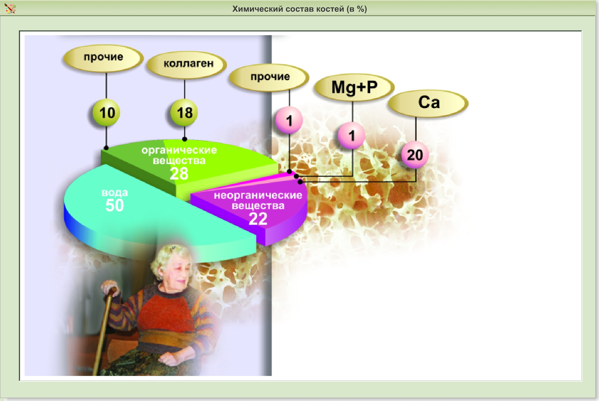
The chemical composition and physical properties of bones
Bone substance consists of mineral salts (about 70%) and organic substances (about 30%). More than half of all minerals are calcium phosphate. The main organics of the bone are proteins collagen and ossein.Minerals give bones hardness and brittleness, organic - flexibility, resilience, elasticity. In general, a combination of organic and inorganic substances gives bones greater strength. The hardness and strength of the bones is comparable to cast iron and brick, so the bones can withstand heavy loads. For example, the tibia tolerates without breaking a load of about 3 tons. The ratio of organic and inorganic matter changes with age. Children have a slightly higher amount of organic matter, so their bones are more resilient, supple and flexible and less likely to break. In elderly and old people, the amount of inorganic substances increases slightly, their bones are less elastic and more fragile, therefore they break more often even with minor injuries.
|
home Shoulder, collarbone, shoulder joint, humerus Elbow joint, bones of the forearm Pelvis, hip Wrist joint, hand Knee-joint Shin, Ankle, Foot Atlas of Anatomy Skeleton Joint prosthetics (arthroplasty) Bone and joint diseases Honey shop Medical sites Feedback |
The influence of internal and external factors on bone development. The skeleton, like any organ system, is part of the body. The development of the skeletal system is influenced by many factors. The influence of internal factors. X-ray examination reveals a number of morphological changes in the bones, depending on the activity of other organs. When radiography is especially clear, the relationship between the skeletal system and the endocrine glands is determined. Active inclusion of the sex glands entails the onset of puberty - the puberty. Before this, in the prepubertal period, the activity of the pituitary gland intensifies. By the beginning of the prepubertal period, all the main points of ossification appear, and there is a gender difference in the timing of their appearance: girls are 1 to 4 years earlier than boys. The onset of the prepubertal period associated with the function of the pituitary gland coincides with the appearance of an ossification point in the pisiform bone belonging to the category of sesamoid bones. On the eve of the puberty, other sesamoid bones also ossify, namely, at the metacarpophalangeal joint of the first finger. The beginning of the puberty, when, in the words of the famous researcher of the endocrine glands, Bidel, “the sex glands begin to play the main melody in the endocrine concert”, manifests itself in the skeletal system by the onset of synostoses between the pineal glands and metaphyses, the very first such synostosis observed in the I metacarpal bone. Therefore, based on its comparison with other data on sexual development (the appearance of terminal vegetation, the onset of menstruation, etc.), the metacarpal synostosis I is considered an indicator of puberty beginning, that is, an indicator of the onset of puberty; in Leningrad residents, 1 metacarpal bones synostosis occurs at the age of 15–19 years for young men and 13–18 years for girls, that is, somewhat earlier. Full puberty also receives a certain reflection in the skeleton: at this time the synostoses of the pineal glands with metaphyses in all tubular bones end, which is observed in women aged 17 to 21 years, and in men at 19 to 23 years. Since the end of the bone growth process ends with the growth of bones in length, it becomes clear why men, "whose puberty is completed later than women, have higher growth in mass than women." Given this connection of the skeletal system with the endocrine and comparing data on age-related features of the skeleton with data on puberty and overall development of the body, we can talk about the so-called bone age.Thanks to this, according to the x-ray picture of some parts of the skeleton, especially the hand, it is possible to determine the age of a given individual or to judge the correctness of the process of ossification in him, which is of practical importance for diagnosis, forensic medicine, etc. Moreover, if the “passport” age indicates the number of years lived (that is, the quantitative side), then the “bone” age to a certain extent indicates their qualitative side. An X-ray examination also reveals the dependence of the bone structure on the state of the nervous system, which, by regulating all processes in the body, performs, in particular, the trophic function of the bone. With enhanced trophic function of the nervous system, more bone tissue is deposited in the bone and it becomes more dense, compact (osteosclerosis). On the contrary, when the trophism is weakened, bone rarefaction is observed - osteoporosis. The nervous system also affects the bone through the muscles, the contraction of which it controls (which will be discussed below). Finally, various parts of the central and peripheral nervous system determine the shape of the surrounding and adjacent bones. So, all vertebrae form the spinal canal around the spinal cord. The bones of the skull form a bone box around the brain and take the form of the latter. In general, bone tissue develops around the elements of the peripheral nervous system, as a result of which bone channels, grooves and fossae appear, which serve to pass the nerves and other nerve formations (nodes). Bone development is also very closely dependent on the circulatory system. The entire process of ossification from the moment the first ossification point appears to the end of synostosis takes place with the direct participation of blood vessels, which, penetrating the cartilage, contribute to its destruction and replacement by bone tissue. In this case, the bone plates are deposited in a certain order around the blood vessels, forming osteons with a central channel for the corresponding vessel. Therefore, the bone, when it occurs, is built around the vessels. This also explains the formation of vascular channels and furrows in the bones at the site of passage and adjoining of arteries and veins. Ossification and bone growth after birth also occur in close dependence on blood supply. As shown by the studies of M. G. Prives, it is possible to outline a series of stages of age-related bone variability associated with corresponding changes in the bloodstream (Fig. 1). one. Neonatal stage characteristic of the fetus (last months of fetal development) and the newborn; the vascular bed of the bone is divided into a number of vascular regions (pineal gland, diaphysis, metaphysis, apophysis) that are not communicated with each other (isolation, isolation) and within the limits of which the vessels are not connected to each other, do not anastomose (the terminal nature of the vessels, “limb” ) 2. Infantile stage, characteristic of children before the onset of synostosis; the vascular regions are still fragmented, but within each of them the vessels anastomose with each other and their terminal character disappears (“isolation”). 3. Juvenile stage characteristic of young men, it begins with the establishment of connections between the vessels of the pineal gland and the metaphysis through the pineal cartilage, due to which the "closure" of the pineal, metaphysical, and diaphyseal vessels begins to disappear. four. Mature stage peculiar to adults; synostoses occur and all intraosseous vessels form a single system: they are not “closed” and not “final”. 5. Senile stage peculiar to old people; vessels become thinner and the entire vasculature is poorer. The shape and position of the bones are also affected by the viscera, for which they form bone containers, the bed, the fossa, etc. The formation of the skeleton and organs refers to the beginning of embryonic life; during their development, they influence each other, which is why the correspondence of organs and their bone containers, for example, the chest and lungs, the pelvis and its organs, the skull and brain, etc. is obtained. In light of these relationships, the development of the entire skeleton should be considered. The influence of external (social) factors on the structure and development of the skeleton. By acting on nature in the course of labor activity, a person sets in motion his natural tools: arms, legs, fingers, etc. In the tools, he acquires new artificial organs that complement and lengthen the natural organs of the body, changing their structure. And man himself at the same time changes his own nature. Consequently, labor processes have a significant impact on the human body as a whole, on its apparatus of movement, including the skeletal system. Especially brightly reflected on the skeleton is the work of muscles. In the places of attachment of the tendons, protrusions (tubercles, processes, roughness) are formed, and at the places of attachment of muscle bundles - smooth or concave surfaces (pits). The stronger the muscles are developed, the better the muscle attachment sites are expressed on the bones. That is why the bone relief due to muscle attachment is more pronounced in an adult than in a child, in men - stronger than in women. Long and systematic contractions of the muscles, as is the case with physical exercises and professional work, gradually cause a change in bone metabolism through the reflex mechanisms of the nervous system, resulting in an increase in bone substance called working hypertrophy This working hypertrophy causes changes in the size, shape and structure of bones, which are easily determined radiographically in living people. In people involved in physical education, the skeleton is much better developed than in people who are not involved. In children of a stronger physique, the skeletal system differentiates much better than in children of a weak physique. Thanks to rational physical measures, the skeleton of children develops better in all departments, including the chest, which has a beneficial effect on the development of vital organs (heart, lungs) contained in it. Therefore, skeletal development data is important for school hygiene. Bone changes due to physical activity are the result of functional conditions. The following facts testify to this. If symmetrical limbs are loaded equally, then the bones on both sides are thickened equally. If the right or left arm or leg is loaded more, then the corresponding bones of the right or left limb are thickened more. Consequently, not only congenital factors (right- or left-handedness) are decisive in the degree of development of bone substance, but also the nature of physical activity after birth throughout a person’s life. This pattern allows through physical exercises to directionally affect bone growth and contributes to the harmonious development of the human body. This, in particular, is the effectiveness of anatomy. Physical therapy, which helps to heal bone injuries, is based on the same pattern. A clear illustration of the role of function in bone formation can be the formation of a pathological joint after a fracture. In case of non-growth of bone fragments, their ends, due to prolonged friction against each other under the influence of muscle contraction, take the form of smooth articular surfaces and the so-called false joint (pseudoarthrosis) forms at the site of the former fracture. Or another example. If a piece of the tibia is transplanted instead of the resected area of \u200b\u200banother, humerus or femur, the transplanted piece of bone (graft) will gradually acquire the structure of the bone (humerus or femur) into which it is transplanted. The architectonics of the transplanted area undergoes restructuring in accordance with the new functional requirements for the transplant. The individual variability of the skeletal system is determined by both biological and social factors. Environmental irritants are perceived by the body biologically and lead to rearrangement of the skeleton. The ability of bone tissue to adapt to changing functional needs through restructuring is the biological cause of bone variability, and the nature of the load, labor intensity, a person’s lifestyle and other social issues are social causes of this variability. Thus, bone is one of the very plastic organs of our body, which undergoes significant changes under the influence of internal and external factors. Many of these changes are detected radiographically, and therefore the x-ray picture of the skeleton becomes a mirror, reflecting to a certain extent the life of the body. Deep study of normal bone structure taking into account working and living conditions is of great importance for solving the issue of the transition of the norm to pathology due to the increased load that goes beyond the norm. This area of \u200b\u200banatomical science is called the anatomy of people of various professions (M. G. Prives) |
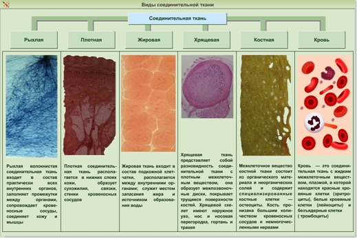
Tissue is a group of cells and intercellular substance that have a common origin, structure and perform similar functions. In the human body there are four main groups of tissues: epithelial, connective, muscle and nerve.
Epithelial tissue form the integument of the body and mucous membranes of internal organs and cavities. The epithelium forms the majority of the glands.
There are several types of epithelium. Stratified skin epithelium and its derivatives: nails and hair, - perform a protective function. Epithelial tissues are composed of closely adjacent cells. The intercellular substance is small. Thus, an obstacle to the penetration of microbes and harmful substances is created. Epithelial cells die in large numbers and are replaced by new ones due to their ability to rapidly reproduce.
A single-layer intestinal epithelium provides the absorption of food digestion products, in the lungs - the absorption of oxygen and the release of carbon dioxide.
The ciliated epithelium in the airways has movable cilia. With their help, dust particles are removed from the respiratory tract.
Connective tissue contains a large amount of intercellular substance.
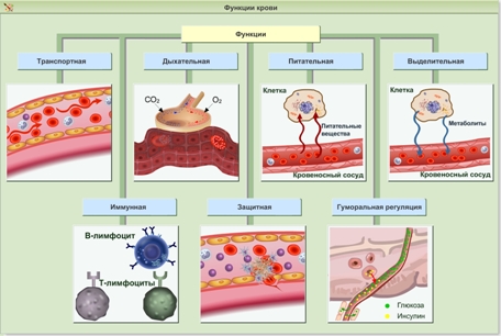
Blood and lymph consist of liquid intercellular substance and blood cells - bind all organs, transferring various substances (connective function); participate in the nutrition of the body (trophic function); cells produce antibodies and carry out phagocytosis (protective function). The flat form of red blood cells and the absence of a nucleus provide a large surface area, which is important for oxygen metabolism. Phagocytes have receptors on the surface that recognize foreign bodies and trigger the process of phagocytosis.
Bone tissue consists of bone plates, inside which cells lie - it has high hardness and forms bones of the skeleton.
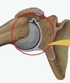
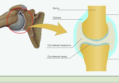
In the cartilage tissue, the intercellular substance is elastic, dense - it is found in the joints, between the bodies of the vertebrae.
The intercellular substance in the form of fibers in dense connective tissue - forms ligaments and tendons (mechanical function).
Adipose tissue is rich in cells filled with fat - storage and protective function (protects from the cold and softens strokes).
Muscle consists of muscle fibers that provide muscle contractions (motor function). Cells of cardiac muscle tissue are connected by special contacts, providing simultaneous contraction of the entire muscle; contain a lot of mitochondria, which is associated with a large load.
Neural tissue it is formed by neurons having processes, which allows for excitation, and neuroglia, which provides protection, support and nutrition of neurons. The long process of the neuron - the axon - reaches a length of 1.5 m and is part of the nerve fibers. The myelin sheath of the axon provides a high transmission rate of the nerve impulse.
Nerve tissue forms the brain and spinal cord, nerve nodes and nerves. It supplies the body with information about the external environment, integrates organs into an integrated organism.
3. How to prevent dental diseases?
- The most important role for dental health is played by proper metabolism. A complete diet containing enough calcium, vitamins, without excess carbohydrates, especially sugar. Vegetables, fruits in the diet. To give up smoking.
- Chewing food, which should not be too soft, creates a load on the chewing apparatus, improves blood circulation and nutrition of teeth.
- It is necessary to rinse your mouth with water after each meal. It is also useful to finish your meal with raw carrots, a solid apple. Stuck food debris with a toothpick or floss.
There is evidence that black tea helps prevent tooth decay.
- Regular brushing, the use of not too hard toothbrushes, scrubbing food debris, without injuring the gums and without erasing the enamel of the teeth. The use of fluoride-containing toothpastes helps prevent tooth decay.
- Visit your dentist regularly every six months for early detection and treatment of tooth decay.
- During meals, you can not alternate hot and chilled foods, which can lead to cracks in the enamel.
- It is not recommended to gnaw hard nuts, bones, open bottles with teeth, etc., bite the thread when sewing, grip small nails in teeth, etc.
- An incorrect bite can develop due to the habit of holding a pen or pencil in your teeth.
- Violations of tooth enamel can occur as a result of the use of certain drugs, for example, tetracycline. Including mother during pregnancy.
"Connective tissue of animals"
TARGET: introduce the features of the structure and functions of the connective tissues of animals.
TASKS:
Educational:
· Find out the location, structure, meaning of connective tissue;
establish the relationship of the structure and functions of the studied tissues;
· To formulate the ability to analyze.
Developing:
· Develop the ability to compare, analyze, generalize;
· Continue work on the formation of the ability to determine tissue from microphotographs;
· Develop communication skills.
Educational:
· Continue work on the formation of a scientific worldview.
PLANNED RESULT: name and identify the cells of the connective tissue of plants, be able to describe them.
BASIC TERMS AND CONCEPTS: fibrous connective tissue, bone tissue, cartilage, blood, plasma, lymph, tissue fluid, adipose tissue
MAIN CONTENT:
1. Cells of mechanical tissue.
2. Cells of conductive tissues - wood and bast. Their location, structure, functions.
Equipment : UMK “Spheres” in biology; definition cards, microphotographs drugs, posters on the topic of the lesson.
Lesson structure:
Organizational moment - 3 min.
Learning new material - 23 min.
Fastening - 10min.
Homework - 2 min.
Reflection - 5 min.
Lesson summary - 2 min.
During the classes:
timeTraining Methods and Tools
I. Organizational moment - 3 min.
Greeting, wording of the topic of the lesson, psychological attitude to work.
Guys, in order for us to work effectively in this lesson, we need to tune in to work. Look at the desk. An epigraph to our lesson is written there. Let's read it in chorus.
“Don't be ashamed not to know
I am ashamed not to study. "
How do you understand this statement?
Do you agree with him?
So let’s take our lesson today under this motto.
In front of you on the tables are colored stickers red, blue, green.
Look at them carefully and choose the one that matches your emotional mood right now. Red color - you are full of energy, ready to work actively. Green color - you are calm. Blue color - you experience a feeling of anxiety, anxiety. Attach it to the edge of the table.
So, let's not waste time and get to work. Record today's number.
Pay attention to the slide from the presentation to the lesson (joint, blood, cartilage)
The theme of our lesson may sound ....
What do you think we will do today in the lesson? (Set the goal of the lesson)
Teacher's introduction
Question to the class
The teacher concludes the psychological state of students.
Creating a problematic situation.
20 - 23 minutes
Learning New Material
Lesson Topic: “Animal Connective Tissue”
Open your textbooks and find the main questions that we have to study today in the lesson:
1What is the connective tissue of animals?
2. What types of connective tissue exist.
3. What are the functions of connective tissue?
In order to make it easier for you to learn new material, recall from the previously studied and answer my questions:
1. What is fabric?
2. What plant and animal tissue do you know?
3. Is the structure and function of the tissue interconnected?
4. What do you think, and in animals, the structure of the tissue will determine their functions?
Of great importance in the life of animals is connective tissue. media objects
Find examples of connective tissue.
Why are such different fabrics combined into one type?
AT output: A common structural feature is that the cells seem to be scattered in the mass of intercellular substance. The structure is the same, but the functions are different.
Connective tissue
Varieties of fabricStructural features
Location
Fibrous
Cartilaginous
Fat tissue
·one. Fibrous connective tissue :
Structure cells are surrounded by a dense network of fibers.
Location -everywhere
Functions - binds the skin to the muscles, holds, interconnects organs.
2. Bone tissue .
What is bone tissue made of?
What combination allows bone tissue to perform a supporting function?
Structural features - solid and strong intercellular substance secreted throughout life. Cells are interconnected by processes.
Location - bones.
Functions - internal support
3. Cartilage
How is cartilage different from bone?
The structural features of tissue cells immersed in an elastic intercellular substance.
Location The skeleton at the junction of bones, the head of the bone, respiratory system (trachea), auricle, tip of the nose.
Functions: giving flexibility to the skeleton
Bone, cartilage, and muscle tissue (we will learn muscle tissue in the next lesson) provide mobility to the body.
Let's take a short break in the lesson and move a bit.
We get up, examine the covers of the desks, take a look under the seats of the chairs. You need to find the components of Voltaire's statement, assemble it and attach it to the board. Forward.
Thank. We have collected the famous statement of Voltaire -
"Movement is life".
4. Blood.
Structural features intercellular substance liquid -plasma.
There are blood cells in the plasma (Platelets, white blood cells, red blood cells)
Location The body of the animal, the blood moves through the vessels.
Functions the binding of various parts of the body into a single organism. Moving through the capillaries, nutrients and oxygen penetrate into the intercellular space.
BLOOD ---- TISSUE LIQUID ------ Lymph
5 . Adipose tissue.
Structural features a large number of fat cells.
Location in the subcutaneous fat layer
Functions deposition of fat reserves, heat preservation and protection against external shocks, water reserve.
Work with the textbook test p. 64.
Frontal conversation with the class on page 64
Teacher's story
Question to the class
Work with keywords.
Teacher's story
Question to the class
Work with keywords.
Work with a textbook, multimedia application, filling out a table during the discussion of the received material.
Work with the textbook on pages 64-65
Teacher's story
Question to the class and filling in the table
Characteristics of each type of connective tissue
The story of the teacher with a phased filling in the table in
Physical education (relaxing pause): for health and emotional relaxation.
Work with keywords
Working with Figure 3.18
Work with keywords.
Working with the drawing Figure 3.19 page 65 of the textbook
10 - 13 min
III. Fastening:
1) Performance of Laboratory work No. 7
"The structure of animal connective tissue"
Connective tissue functions
Subcutaneous tissue, between internal organs
Stocking, protective,
water source
Cavities of the heart and blood vessels
Respiratory, transport, protective
2) Perform test items in worksheets
Be careful and complete each task independently. I wish you success!
1 Adipose tissue functions DO NOT:
A) storage of substances;
B) heat preservation;
C) ensuring the flexibility of the body;
Damage protection
2. Complete the phrase
"The blood contains a liquid intercellular substance-- _________-
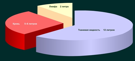
3. Choose the most complete and correct answer. The internal environment of the living is:
B) blood and lymph;
C) blood, lymph and tissue fluid;
D) blood, lymph, tissue fluid and water.
4. Select the correct statement:
A) tissue is a group of cells with a common structure
B) tissue is a group of cells with a similar structure.
C) tissue is a group of cells that perform a specific function.
D) tissue is a group of cells of the same size.
5. General feature of the structure of connective tissue of animals:
A) cell formation;
B) damage protection
C) cells are scattered in the mass of intercellular substance
D) the formation of new cells.
Answers: 1 - A, B. 2- plasma. 3-B. 4- B.
Have you completed the test? I ask you to check the correctness of this task from each other Checked?
Set a rating based on the following parameters:
“5” - everything is correct (100%)
“4” errors (80 - 60%)
“3” - 3 errors (50 - 30%)
“2” - 4 or more errors (less than 20%)
3) Work in a notebook exercise machine .
Notebook-practical page 20-23
Perform laboratory work №7 step by step
Fill out the final table using a slide or tables in a notebook.
Make a conclusion about the relationship between the structure and functions of different types of connective tissue
Check the correctness of filling the table
Performing the test on sheets (one remains with the student for self-assessment, the second is then checked by the teacher to verify the objectivity of the assessment)
Work in pairs "mutual check"
Accomplishment of a task in a notebook simulator p. 24.№ №17,18,19, p. 29 №4
IV. Homework (Differentiated)
- § 24 p. 64-65; orally, learn the terms
- § 24, answer questions p. 65; in the exercise book
Page 30 No. 2, Pages 31 No. 4.
To prepare a message on the topic “Blood Its Role in the Body” and “The Importance of Adipose Tissue in the Life of Animals” (see “Reading Book” on the electronic application), task at will.
Writing homework on the blackboard and in the diary.
V. Lesson summary
Let us return to the questions that were posed at the beginning of the lesson. Did we answer them?
The teacher leads students to the conclusion of the lesson:
In living organisms, the relationship of the structure and their functions is traced.
Grading all students, taking into account individual work, frontal work during a conversation, assessment on the test.
Question to the class.
Lesson wording.
VI. Reflection -
Reflection Issues:
Was an electronic application necessary to explain the material, or could you study this topic using only a textbook?
Did you like today's lesson? Analyze the lesson and fill out the table as honestly as possible.
Learned in the lesson
I want to know
Choose from stickers the one that matches your emotional mood right now. Glue it to the edge of the desk.
In conclusion, invite the guys to thank each other for successful cooperation with a solemn handshake.
Thank you all for the lesson. The lesson is over.
Conclusion about the emotional state of students after class
1 Discover the relationship of the structure and basic functions of human tissues?
Answer:
a collection of cells and intercellular substance, similar in origin, structure and adapted to perform certain functions, called tissue. Four main types of tissues are distinguished in the human body: epithelial, connective, muscle and nervous. Epithelial tissue forms a layer of cells that make up the mucous membranes of all internal organs and body cavities. Through the epithelium, a metabolism occurs between the body and the environment. In the epithelial tissue, the cells adhere closely to each other. The intercellular substance, as a rule, is undeveloped. Due to this, a barrier is created for the penetration of microorganisms, harmful substances into the body; reliable protection of tissues located under the epithelium is provided. There are various types of epithelium depending on the structure of cells: squamous epithelium, cubic, cylindrical, glandular and ciliary. Each type of epithelium lines certain organs and performs a characteristic function. For example, the glandular epithelium fills the secretory organs, the ciliary epithelium lines the nasal cavity, thereby preventing the movement of cilia, the penetration of dust and other objects into the internal respiratory organs. A feature of the connective tissue is the strong development of intercellular substance. Connective tissue includes blood, lymph, cartilage, bone and adipose tissue. The main functions of connective tissue are trophic (food) and supporting. Blood and lymph are liquid connective tissues, which, carrying out the transfer of substances throughout the body, provide nutrition, respiration, immunity of tissues, organs and the relationship between organs. Fibrous connective tissue consists of cells interconnected by an intercellular substance in the form of fibers. Fibers can lie tight and loose. Fibrous connective tissue is present in all organs. Adipose tissue containing many fat-filled cells is similar to loose connective tissue. In the cartilage tissue, the cells are large, the intercellular substance is elastic, dense, contains elastic and other fibers. There are many in the joints, between the bodies of the vertebrae. Bone tissue consists of bone plates, inside of which lie cells connected to each other by many thin processes. Bone tissue is hard. Muscle tissue is formed by individual cells - muscle fibers, which contain the finest contractile fibers - myofibrils. The latter has such a name because its fibers have a transverse striation due to the correct alternation of light and dark discs. Cross-striped muscle tissue is often divided into skeletal and cardiac. Skeletal consists of elongated fibers up to 10-12 cm long and provides a movement function. Cardiac muscle tissue, like skeletal, has a transverse striation, but unlike skeletal, there are special areas where the muscle fibers are tightly closed. Due to this structure, the contraction of one fiber is quickly transmitted to others, providing simultaneous contraction of a large section of the muscle. The walls of the internal organs — the stomach, intestines, bladder, and blood vessels — are built from smooth muscle tissue. Smooth muscles regulate their contractions and changes in the diameter of blood vessels. Nerve tissue performs the functions of perception, processing, storage and transmission of information coming from both the environment and from the inside of the body. The activity of the nervous system provides the body's response to various irritations and coordination of the work of various organs of animals and humans. Nerve cells - neurons usually have a stellate or spindle-shaped form and consist of a body and processes (axon and dendrites). The sheathed processes of nerve cells are called nerve fibers. The main properties of a neuron is the ability to be excited and conduct impulses along nerve fibers. Branched processes (dendrites) conduct excitation to the body of the neuron, and one long process (axon) - from the body of the neuron. In most cases, neurons are located in the nerve centers - the brain, ganglia and nerve nodes. Nerve tissue is part of the body as part of it and provides a combination of functions of all other parts of the body. Each tissue consists of cells with a certain shape, size, function. Morphofunctional integrity of the whole organism is achieved only through the interaction of all tissues.
Related questions
- A rectangular section, whose width is 60 m and a length of 30 m more, is surrounded by a metal mesh 1.6 m high. Find the area of \u200b\u200bthe metal mesh. How much they paid for the fence, if 1 sq.m. a fence costs 800 rubles?
Ticket number 1
- Compare the structure of plant and animal cells, as evidenced by their similarity?
A cell is the main structural, functional and reproducing element of a living organism. Common signs:
1. The unity of structural systems - cytoplasm and nucleus.
2. The similarity of metabolic processes and energy.
3. The unity of the principle of the hereditary code.
4. Universal membrane structure.
5. The unity of the chemical composition.
6. The similarity of the process of cell division.
| Plant cell | Animal cage | |
| Size (Width) | 10 - 100 microns | 10 - 30 microns |
| The form | Monotonous - cubic or plasma. | The form is various |
| Cell wall | The presence of a thick cellulose cell wall is characteristic, the carbohydrate component of the cell membrane is strongly expressed and represented by cellulose cell wall. | As a rule, they have a thin cell wall, the carbohydrate component is relatively thin (thickness 10 - 20 nm), it is represented by oligosaccharide groups of glycoproteins and glycolipids and is called glycocalyx. |
| Cell center | In lower plants. | In all cells |
| Core position | The nuclei in highly differentiated plant cells, as a rule, are squeezed by cell sap to the periphery and lie parietally. | In animal cells, they most often occupy a central position. |
| Plastids | Characteristic for cells of photosynthetic organisms (photosynthetic plants - organisms). Three main types are distinguished depending on color: chloroplasts, chromoplasts and leukoplasts. | no |
| Vacuoli | Large cavities filled with cellular juice - an aqueous solution of various substances that are reserve or final products. Osmotic cell reservoirs | Contractile, digestive, excretory vacuoles. Usually small |
| Inclusions | Spare nutrients in the form of starch grains, protein, drops of oil; vacuoles with cell juice; salt crystals | Spare nutrients in the form of grains and drops (proteins, fats, glycogen carbohydrate); end products of metabolism, salt crystals; pigments |
| Division method | Cytokinesis by the formation of a fragmoplast in the middle of a cell. | Division by the formation of constriction. |
| Power way | Autotrophic (phototrophic, chemotrophic) | Heterotrophic |
| Ability to photosynthesis | there is | no |
2.Explain the need for environmental protection.
As a result of environmental pollution, global environmental problems arise, their presence and there are examples of the need to protect the environment:
1. acid rain
2. ozone "holes"
3. deterioration of human health, the occurrence of hereditary diseases
4. death of animals and plants, a decrease in the number of their species on Earth, a change in biodiversity
5. change in the habitat of organisms and change of ecosystems
6. The decrease in freshwater reserves on the planet
7. The problem of waste disposal (domestic and industrial)
8. the occurrence of a “greenhouse effect”.
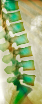
3. What causes spinal curvature and flat feet, how to prevent them?
Curvature of the spine - changes in the normal configuration of the spine. The spine of an adult properly folded person has characteristic bends: in the cervical region, the spine is bent forward (cervical lordosis), in the thoracic region forward (lumbar lordosis) and in the sacral region - backward. . Side bends
there is no normal spine. Spinal curvatures can occur in cases of intrauterine skeletal development disorders - the formation of wedge-shaped and additional vertebrae,
incorrect formation of the 5th lumbar vertebra and ribs, etc. This is the so-called. congenital curvature. Sometimes it arises as a result of some disease (rickets, polio, tuberculosis, etc.), injuries (spinal fractures), with disturbances in proper standing, with different lengths of the lower extremities, etc. At a later age, after the formation
skeleton, curvature of the spine develop in clerical workers, violinists, shoemakers, etc., whose work is associated with a long stay in one position. The muscular system plays an important role in their formation. With the development of spinal deformity, the uniform traction of the muscles surrounding the spine is disrupted, which in turn exacerbates the existing curvature.
Under flat feet mean flattening of the arches of the foot. Due to the resulting deformation, the muscles, and then the ligaments of the legs, weaken. When walking, pains are felt in the foot, ankle joint, lower back. In 10% of patients, pathology is detected at birth. In other cases, the disease is acquired throughout life.
Causes of spinal curvature and flat feet:
1. Too wide or narrow shoes without heels;
2. excessive load on the foot;
3. excessive joint flexibility;
The main principle of maintaining proper posture is prevention. The experience of orthopedic specialists convinces us that the main role in the formation and maintenance of correct posture belongs to upbringing and systematic physical exercises. Useful skills are easily developed in childhood, therefore, it is necessary to begin to form the correct posture before school: Tables and chairs should correspond to the child’s height. You need to teach children to stand, sit and not slouch while walking from the age of 4. Moderate cold wiping will not only harden the child’s body, but will also increase muscle tone. An important role is played by proper nutrition with a sufficient content of all the necessary substances - proteins, carbohydrates, fats, vitamins and trace elements.
Flatfoot Warning:
· It is necessary to pay special attention to shoes in children;
· Special exercises, also try to walk barefoot more.
Ticket number 2
Discover the relationship of the structure and basic functions of human tissues.
the cloth - a system of cells and intercellular substance, united by a common origin, structure and functions. In the human body there are four main types of tissues: epithelial, connective, muscle and nervous.