You are interested in how your internal organs work, how they perform various functions necessary for a fulfilling life?
Of course, we are still at school in biology lessons about the human body. Remember:
Skeleton - protects our internal organs and determines the shape of the body.
Internal organs are constantly busy with something, they are not used to resting.
human anatomy is a science that studies the origin, development, forms and structure of the human body. Anatomy studies both the external forms and proportions of the human body and its parts, as well as individual organs, namely their structure and microscopic structure. Human anatomy is closely related to physiology - the science of the vital functions of the body and organs. Without knowing the structure of an organ, it is impossible to understand the causes of its disease. Without knowing the functions that each organ system performs, it would be impossible not only to treat the simplest diseases, but even to make a diagnosis.
Human anatomy clearly demonstrates how completely and at the same time vulnerable the human body is, where damage to one organ can lead to the failure of all organ systems in the body.
1. Structure of a human cell
The human body is very complex. According to its biological characteristics, it ranks among the higher animals. But man as a whole is not an animal; the usual measurements adopted by zoologists cannot be applied to him. Man has hands, and the brain has reached such a high degree of development that it allows him to perform extraordinary complex species work and truly rule the world.The most common elementary form of organization of living matter is the human cell. This carrier of life gave rise to the development of all plant and animal life on earth. It became entrenched in evolution and became part of complex multicellular organisms.
 The structure of a human cell.
A small lump of protoplasm with a nucleus is what a cell is. Its dimensions are measured in hundredths of a millimeter. At the same time, it is already an extremely complexly organized living system. It has its own elements (organelles), its own distribution patterns nucleic acids and protein molecules, although in relation to the whole organism the cell is just a small structural unit.
There are a huge number of cells in the human body. They are varied in appearance and are different in their properties. Thus, muscle cells capable of contracting have an elongated or spindle-shaped shape, and nerve cells, whose task is to transmit excitations and carry out communication functions, have long processes.
But a cell in the human body can only live within tissues. 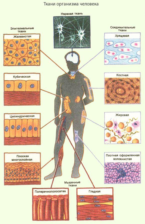
2. Human tissue
A cell in the human body can only live within tissues. Cells that are homogeneous in structure and unambiguous in function, originating from a common rudiment, form tissue that performs more complex tasks. In accordance with the history of development, with the laws structural organization and participation in performing a specific job, all the variety of tissues comes down to four main types.Types of human tissue:
. epithelial
. muscular
. nervous
. tissues of the internal environment.
Functions of human body tissue
Pokrovny epithelium is located either on the surface of the skin or on the surface of the membranes lining the internal hollow organs and called mucous membranes. Another type of epithelium, glandular epithelium, is used to build glands, special organs that produce chemicals needed by the body.
Tissues of the internal environment constitute a variety of connective tissue. This includes all types of supporting tissue - bone, cartilage, dense connective tissue, which we find in the periosteum, in ligaments, in muscle tendons.
Another group of tissues of the internal environment are trophic tissues - blood, lymph, loose connective tissue, adipose, pigment, etc. Characteristic feature All these tissues are characterized by the presence in them, along with cells, of intercellular living substance. It is presented either in a form - fibers, or in the form of an amorphous base substance.
Functions of human tissue.
From muscle tissue We will describe two separately.
Smooth muscle tissue built of smooth muscle spindle cells - fibers. It makes up the muscular layer internal organs and vessels. Contraction of smooth muscle fibers occurs regardless of our desire, under the influence of internal, not subordinate to consciousness, reasons.
Structural element striated muscle tissue consists of fibers with a large number of nuclei, longitudinally located myofibrils (thin filaments enclosed in protoplasm - it is due to them that the fiber contracts) and transverse striation, which is explained by the alternation of light and dark discs along the length of the fiber. All skeletal muscles of the body, subordinate to our will, are built from striated muscle tissue.
Nervous tissue differs in its cellular and fibrous structure. Its structural elements are nerve cells and glia cells that accompany the nerve elements.
Organs are formed from tissues. It's more high shape organization of the internal structure of the body. The participation of a number of organs in the performance of any single, albeit multi-stage work allows them to be combined into organ systems. For example, the digestive system, the respiratory system, etc. The bone, joint and muscle systems form the movement apparatus.
![]()
3. The structure of the human spine.
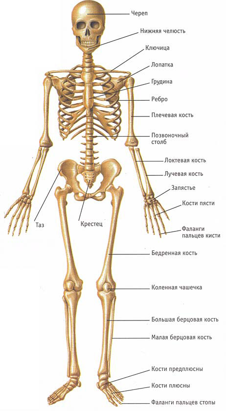
The human skeleton and its supporting structure can truly be called an engineering marvel of Mother Nature. More than two hundred bones, connected in a bizarre but very rational way, play the role of a frame, an armature of our body. The human skeleton is a unique protection of the entire body from stress and damage to vital organs and tissues. Our brain is protected on all sides by the bones of the skull, the heart, lungs, liver and esophagus - by the chest, reproductive organs and bladder- pelvic bones.In order for the body to be mobile, nature created the ligamentous apparatus, and to avoid unnecessary friction and wear in places of heavy load, joints appeared. Muscles are attached to the bony base of the skeleton, the task of which is to direct the movement of the limbs and individual parts of our skeleton.
The spine itself, or, as they still not very affectionately say, the ridge, is the basis of our skeleton. But you and I are unlikely to realize how complex the system the spinal column itself is. Many bones are connected to each other by ligaments, covered by muscle groups, but at the same time they not only retain sufficient mobility, but also take on a huge load. All the abundance of our possibilities for movement in space is provided the most complex system bones and ligaments, cartilage and small joints.
What exactly allows us to maintain motor activity under very heavy loads and not fall apart or experience pain? The answer to this question is very, very simple - the structure of our spine, a special system of ligaments, cartilage and vertebrae.
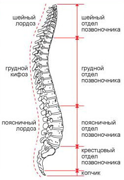
The spinal column consists of 32 vertebrae, which, depending on their shape and position, are divided into cervical, thoracic, lumbar, sacral and coccygeal. The sacrum and coccyx are formed not by individual vertebrae, but by conglomerates of vertebrae fused together, which have lost mobility, but are capable of bearing a huge load.
In the cervical spine there are 7 vertebrae, the thoracic spine consists of 12 vertebrae, the lumbar spine - of 5. Thin, almost openwork vertebrae of the cervical spine pass into more massive, dense thoracic vertebrae, which, in turn, rest on large, thick and very powerful lumbar vertebrae. These sections represent a mobile part of the spine, in contrast to the underlying fixed sacrum and sedentary coccyx.
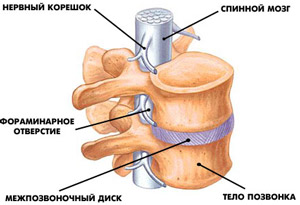 But the vertebrae don't just lie on top of each other. They are connected to each other using intervertebral discs, which simultaneously connect the vertebrae to each other and ensure their mobility. Intervertebral discs consist of two parts: the so-called core, which includes special cartilage and water, and the surrounding ring of dense connective tissue fibers. The core of the intervertebral disc takes on the main load falling on the spinal column, and the dense ring prevents the core from being flattened by pressure, supporting it on all sides.
In addition, the vertebrae are held together by spinal ligaments - long ones that run along the entire spine along its anterior and posterior surfaces, as well as short ones that fasten individual vertebrae together. Ligaments connect bones - vertebrae, located around small joints and the vertebrae themselves. Connective tissue fibers are also attached between the intervertebral disc and the vertebral body. Thus, the integrity of the entire spine is ensured by strong, reliable ligaments.
 The mobility of the spine, the ability to turn, bend to the sides, flexion and extension are provided by numerous joints, both between the vertebrae and between other elements of the skeleton, such as ribs. The joints of the spine are formed by the processes of the vertebrae; they have a small range of movements in each individual joint, but in general they a large number of allows you to make tilts and turns with a fairly wide amplitude.
Along the entire spine, muscles lie in comfortable spaces. They determine our ability to move in space and form the gait, posture and figure unique to us. Muscles are formed by many thin fibers, each of which acts like a tourniquet - lengthens when stretched and shortens when compressed.
Muscles form the muscular corset of the body, including the muscular corset of the spine. They are located in separate groups, among which there are small and large muscles, and, for example, at the level of the spine, the muscles are located in 8 layers. One muscle is located above the other, and thus a very dense “sandwich” is formed, reliably covering the bones and ligaments.
The next component of the spinal column necessary for our full life can be considered nervous tissue and the spinal cord. The spinal cord is located inside the spine and is securely covered by bone tissue. Nerve trunks emerge from the spinal cord, providing the vital functions of our entire body, from the top of the head to the toes. Heat, cold, touch, pain, pleasure and disgust - all these reactions to external and internal stimuli are provided by the nervous system.
The nervous system is part of our movement, passing through its channels the necessary impulses to specific muscles working at the moment. In addition, the nervous system controls the functioning of all our internal organs - the liver and heart, lungs, kidneys, etc.
This fact is related to the statements of many doctors involved in the treatment of spinal diseases that certain manipulations with the back can cure asthma or stomach ulcers. As you can see, there is some truth in this, since pinched nerves perform their functions poorly and cannot transmit the correct signals, which causes the disease.
Of course, Nature very skillfully combined all the individual parts of the spine into a single whole and thus allowed us not just to live, but to live actively, in motion, moving in space. After all, from a biology course we know that all life on earth is divided into two large groups: invertebrates and chordates, that is, having a core - a chord or spine.
There is no doubt that vertebrates are at a higher level evolutionary development, as they have greater chances of survival. And in humans, to these factors, bipedalism is also added - such distinguishing feature. Of course, it immediately affected the structure of the spine, its anatomy and physiology. The lower lumbar vertebrae became denser and noticeably thicker, the anterior ends of the ribs sank, and the former tail fused together to form the tailbone.
 But this was not enough for Nature, and she created several bends in our spine that help us withstand a fairly large load. Our spine has become like a spring with several depressions and bumps. It expanded from below and became powerful, the sacrum firmly “hooked” onto the pelvic bones to allow us to stand and walk reliably.
To this day, the human spine follows these curves. They begin to form in children from a very young age. First, the child raises his head - a cervical curve is formed, then the baby sits down and bends in the thoracic region, and later, when the little man learns to walk, a lumbar curve of the spine forms. Nature reasoned very simply: more load means more bends, and our spine is a wonderful example of this rule.
Doctors named these curves using Latin and Greek words - kyphosis and lordosis. Kyphosis is any backward bend of the spine, and we have two of them: large in the thoracic region and small in the sacral region. Despite the fact that the bones of the sacrum are fused together, they are still not completely flat, but are curved back and form an arch, similar to the tail of a frightened animal. The medical term lordosis is any forward bend of the spine, and we also have two lordoses: large in the lumbar region and small, but very mobile, in the cervical region.
Which doctors should I contact for a Spine examination? Traumatologist, Orthopedist, Vertebrologist, Physiotherapist.
What diseases are associated with the Spine: Lumbar spinal stenosis, Ankylosing spondylitis, Spinal osteochondrosis, Low back pain, Intervertebral disc herniation, Scoliosis.
What tests and diagnostics need to be done for the Spine: MRI of the spine, CT scan of the spine, X-ray of the spine.
Well, these are probably all the structural features of the human spine that you need to know in order to understand how his diseases are formed and, most importantly, how to prevent and treat them.
4. Secrets of the joints and ligaments of the spineWe wouldn’t be able to move at all if it weren’t for nature’s brilliant “invention” - joints. In the human body there are a huge number of different kinds of joints and joints, thanks to which the bones have the ability to change their position relative to each other.And since the articular surfaces are covered with a thin layer of cartilage with a smooth surface, they slide very easily against each other. In addition, the area where the bones meet to form a joint is enclosed in a bursa, or joint capsule, which produces fluid to lubricate the joint, further reducing friction. Moreover, the more the joint works, the more this “lubricant” is produced. And another amazing property of joint fluid is that it changes its viscosity depending on the load and ambient temperature. With greater load, for example, if you lift a barbell, the viscosity increases and the joint acquires shock-absorbing properties. Such changes in viscosity values can occur with incredible speed. Among the many joints of the human skeleton, almost none are completely identical. And yet, it is customary to classify joints precisely by their similarity to each other, distinguishing between spherical, ellipsoidal, block-shaped, cylindrical and flat joints. Having mentioned this classification (rather for general erudition), we will not describe the joints in detail here. With one exception - the joints of the spine, on which the flexibility of our entire body depends. |  The structure of the joints and ligaments of the spine |
Have you ever wondered “why is a person taller in the morning?” For some, such growth fluctuations reach 6 cm. This occurs because during the night the intervertebral discs, without experiencing pressure, “rested” and moved the vertebrae apart.
The thicker and more flexible the intervertebral discs are, the more flexible your spine and its parts are. The lumbar region is the most mobile: the thickest intervertebral discs are located here. And the thinnest of them are in the middle part of the thoracic region - here the mobility of the vertebrae is the least. In the cervical spine, the discs are also quite thin, but the height of the vertebrae here is much smaller, so the flexibility of the neck is almost the same as that of the lumbar spine.
Indicators of flexibility of the spine and its parts may differ markedly in different people. With little flexibility, the angular displacement of the vertebrae is regulated mainly by ligaments running along the spine. For those who have significant flexibility, the muscles of the trunk, which are more extensible, come into play.
You need to train your spine flexibility very carefully. This complex biomechanical organ is easily damaged by too intense exercise. But the spine contains many vital centers of our body.
Ligaments
Ligaments (fibrous bundles surrounding the joint) ensure a certain position of the bones relative to each other. At the same time, this kind of soft “reinforcement”, due to its elasticity, allows the joint to move quite freely. With moderate flexion or extension of a particular joint, the ligaments do not strain at all; if the range of movements reaches extreme values, the ligaments begin to stretch, limiting their amplitude. However, the braking capabilities of the ligaments are very limited, so if they are suddenly stretched, you risk injury.
The fact is that the tissue of ligaments and tendons includes collagen and elastic fibers. The first of them provide the strength of the ligaments, and the second, as the name implies, provides elasticity. Moreover, there are more collagen fibers in tendons and ligaments than elastic ones. (True, the content of collagen and elastin in the ligamentous tissue may differ markedly from person to person, depending on the characteristics of the constitution and age. This, in particular, determines the greater or lesser “initial” flexibility of each person.)
So, we found out: the joint is hidden in the articular capsule, entangled with ligaments, which allows it to move. However, by itself it is still helpless and cannot provide movement - this requires the application of external forces. The muscles act as such an external force in relation to the joint. They have the ability to change their length, that is, to contract (we are talking about striated muscles), and to move the joints. At the same time, the muscles limit the range of this movement. The degree of limitation depends on the individual properties of the muscles - their elasticity and extensibility. And the elasticity of muscles, their ability to stretch, is subject to targeted training.
Which doctors should I contact to examine the Ligaments: Traumatologist, Rehabilitologist.
What diseases are associated with Ligaments: Ligament and muscle ruptures, Ligament sprains.
What tests and diagnostics need to be done for Ligaments: Examination by a traumatologist.
Structure and functions of the skin
The skin forms the general covering of the body, protecting the body from external influences. This is the most important organ of the body, performing a number of essential functions: heat regulation, secretion of secretions (sweat and sebum), and with them harmful substances, respiration (exchange of gases), depot of energy reserves. It is also credited with endocrine properties. The main function of the skin is to perceive various environmental irritations (touch, pressure, temperature and harmful irritations). Thus, the skin is a complex complex of perceptive devices with a huge reception surface, reaching an area of about 1.6 m2 in adults.Skin covers the entire human body. In the area of the mouth, nose and other openings it passes into the mucous membrane. Its thickness is not the same. It reaches its greatest development in the center of the back (thickness up to 1 cm).
Skin color depends on chemical substance- pigment deposited in the cells of the outer layer, depending on the degree of development of blood vessels in the deep layer, as well as on its thickness and density. In children, the skin is thinner; blood vessels showing through it give it a pink color. Old people have rougher skin. It thickens, becomes wrinkled, and acquires a yellow or brown tint. Redness of the skin depends on the dilation of blood vessels. Severe nutritional disorders often lead to changes in the natural color of the skin. Darkening of the skin of patients with disorders of the adrenal gland is known. In alcoholics, the red skin of the face and especially the nose is explained by frequent rushes of blood to the vessels of the face and the loss of their elastic properties.
Outer layer skin is called the epidermis (epidermis - cuticle (epi - above, dermis - skin). It is built by several rows of epithelial cells. Superficial cells become keratinized and sloughed off. Their place is taken by new ones, formed by the multiplication of cells of the germinal zone lying in the depths. Such renewal of surface cells continues throughout life. It has a beneficial effect on the protective functions of the skin. Microbes and the toxic substances they produce do not cause pathological painful phenomena when exposed to intact skin. The keratinization of the epidermis is accelerated by chronic friction and pressure (calluses).
Inner, deep layer- the skin itself, or dermis. It is built of dense connective tissue, in which numerous strands of fibers are intertwined, running in different directions. Beneath the dermis lie fascia and fat cells. The skin is connected to them by fibers, vessels and nerves. The dense dermis passes into the subcutaneous fat. 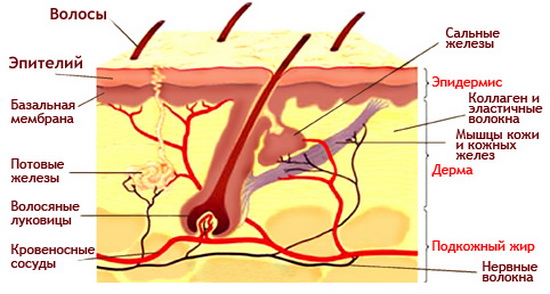
The structure of human skin (diagram).
The outer surface of the dermis is uneven due to the many papillae. The thickness of the dermis is penetrated by vessels, nerves and excretory ducts of the sweat glands. The sweat glands themselves (almost microscopic in size) are immersed in the dermis. During the day, they release up to 600 cubic centimeters of water, and in hot weather and during intense work, much more. Together with the secretions of these glands, excess fluid with salts dissolved in it, as well as waste, ballast substances, are removed from the body.
Another type of skin gland is the sebaceous gland. They are located near the roots of the hair. The secretion product of these glands serves as a lubricant for the skin and prevents hair from becoming brittle. All human skin, with the exception of transitional areas (for example, the red border of the lips), soles and palms, is covered with hair. But it is necessary to distinguish between two types of hair - some in the form of a fluff, never reaching a significant length, others, thicker, grow on the head, surround the genitals, are found in the armpit, on the eyelids. The color of the hair depends on the amount and type of pigment contained in the hair shaft. The disappearance of pigment with age and its replacement by air bubbles leads to the appearance of gray hair. An unresolved problem of our time is still premature hair loss (baldness).
In addition to hair, skin appendages also include nails - hard horny outgrowths of the epidermis. They serve to protect your fingertips.
Taking care of the cleanliness and safety of your skin is taking care of your health. Regular washing hot water leads to the removal of gland secretion products, excess sweat, and dust particles deposited on the skin. The exit openings of the glands and the pores of the skin restore their permeability and can again participate in excretory, respiratory and heat-regulating functions.
Washing and wiping the body with cold water helps to harden the body and has a beneficial effect on the nervous system and the functioning of the heart.
One more protective function of the skin should be pointed out. Under conditions of direct exposure to sunlight, the body receives an increased dose of ultraviolet radiation, which is not always harmless to humans. Cells of the epidermis irradiated by sunlight accumulate pigment grains that block ultraviolet rays and do not allow them to penetrate deep into the body. A tan forms. Its usefulness on a moderate scale is beyond doubt. But for those who do not tolerate heat well, abundant sun rays are undesirable.
Which doctors should I contact for a skin examination? Dermatologist, Venereologist, Oncologist, Cosmetologist.
What diseases are associated with the Skin: Diseases of the skin and subcutaneous tissue.
What tests and diagnostics need to be done for the Skin: Dermatological examination of the skin.
Materials used from the site fiz-ra.com
Next time we will talk about human organ systems
human anatomy tissue
To the article HUMAN ANATOMY The structural and functional unit of living things is the cell - the anatomical basis of most organisms, including humans. Complexes of specialized cells, characterized by a common origin and similarity in both structure and functions, are called tissue. There are four main types of tissue: epithelial, connective, muscle and nervous. See also HISTOLOGY. Epithelial tissue covers the surface of the body and the cavities of various tracts and ducts, with the exception of the heart, blood vessels and some cavities. In addition, almost all glandular cells are of epithelial origin. Layers of epithelial cells on the surface of the skin protect the body from infection and external damage. The cells lining the digestive tract from the mouth to the anus have several functions: they secrete digestive enzymes, mucus, and hormones; absorb water and digestive products. The epithelial cells lining the respiratory system secrete mucus and remove it from the lungs along with the dust and other foreign particles it traps. In the urinary system, epithelial cells carry out excretion and reabsorption (reabsorption) various substances in the kidneys, and also line the ducts through which urine is excreted from the body. Derivatives of epithelial cells are human germ cells - eggs and sperm, and the entire path that they pass from the ovaries or testes (genitourinary tract) is covered with special epithelial cells that secrete a number of substances necessary for the existence of the egg or sperm. Connective tissue, or tissues of the internal environment, is represented by a group of tissues diverse in structure and function that are located inside the body and do not border either the external environment or the cavities of organs. Connective tissue protects, insulates and supports body parts, and also performs a transport function within the body (blood). For example, ribs protect organs chest, fat serves as an excellent insulator, the spine supports the head and torso, and blood carries nutrients, gases, hormones and waste products. In all cases, connective tissue is characterized by a large amount of intercellular substance. The following subtypes of connective tissue are distinguished: loose, adipose, fibrous, elastic, lymphoid, cartilaginous, bone, and blood. Loose and fatty. Loose connective tissue has a network of elastic and elastic (collagen) fibers located in a viscous intercellular substance. This tissue surrounds all blood vessels and most organs, and also underlies the epithelium of the skin. Loose connective tissue containing large numbers of fat cells is called adipose tissue; it serves as a storage site for fat and a source of water formation. Some parts of the body are more likely than others to store fat, such as under the skin or in the omentum. Loose tissue also contains other cells - macrophages and fibroblasts. Macrophages phagocytose and digest microorganisms, destroyed tissue cells, foreign proteins and old blood cells; their function can be called sanitary. Fibroblasts are primarily responsible for the formation of fibers in connective tissue. Fibrous and elastic. Where a resilient, elastic, and strong material is needed (for example, to attach a muscle to a bone, or to hold two bones in contact together), we typically find fibrous connective tissue. Muscle tendons and joint ligaments are built from this tissue, and it is represented almost exclusively by collagen fibers and fibroblasts. However, where a soft, but elastic and strong material is needed, for example in the so-called. In the yellow ligaments - dense membranes between the arches of adjacent vertebrae, we find elastic connective tissue, consisting mainly of elastic fibers with the addition of collagen fibers and fibroblasts. Lymphoid tissue will be discussed when describing the circulatory system. Cartilaginous. Connective tissue with dense intercellular substance represented by either cartilage or bone. Cartilage provides a strong but flexible framework for organs. The outer ear, nose and nasal septum, larynx and trachea have a cartilaginous skeleton. The main function of these cartilages is to maintain the shape of various structures. The cartilaginous rings of the trachea prevent its collapse and ensure the passage of air into the lungs. The cartilage between the vertebrae makes them move relative to each other. Bone. Bone is a connective tissue whose intercellular substance consists of organic material (ossein) and inorganic salts, mainly calcium and magnesium phosphates. It always contains specialized bone cells - osteocytes (modified fibroblasts), scattered in the intercellular substance. Unlike cartilage, bone is penetrated by a large number of blood vessels and a number of nerves. On the outside it is covered with periosteum (periosteum). The periosteum is the source of osteocyte precursor cells, and restoration of bone integrity is one of its main functions. The growth of limb bones in length in childhood and adolescence occurs in the so-called. epiphyseal (located at the articular ends of the bone) plates. These plates disappear when the bone stops growing in length. If growth stops early, short dwarf bones are formed; if growth continues longer than usual or occurs very quickly, we get long bones giant. The rate of growth in the epiphyseal plates and bone in general is controlled by pituitary growth hormone. See also BONE. Blood is connective tissue with a liquid intercellular substance, plasma, making up slightly more than half of the total blood volume. Plasma contains the protein fibrinogen, which, when in contact with air or when a blood vessel is damaged, forms a fibrin clot consisting of fibrin strands in the presence of calcium and blood clotting factors. The clear yellowish liquid remaining after the clot forms is called serum. Plasma contains various proteins (including antibodies), metabolic products, nutrients (glucose, amino acids, fats), gases (oxygen, carbon dioxide and nitrogen), various salts and hormones. On average, an adult male has approx. 5 liters of blood. Red blood cells (erythrocytes) contain hemoglobin, an iron-containing compound that has a high affinity for oxygen. The bulk of the oxygen is carried by mature red blood cells, which, due to their lack of a nucleus, do not live long - from one to four months. They are formed from nuclear cells of the bone marrow and are destroyed, as a rule, in the spleen. There are about 4,500,000 red blood cells in 1 mm3 of blood for a woman, and 5,000,000 for men. Billions of red blood cells are replaced with new ones every day. In the inhabitants of high mountainous regions, the content of red blood cells in the blood is increased as an adaptation to lower concentrations of oxygen in the atmosphere. The number of red blood cells or the amount of hemoglobin in the blood is reduced in anemia (see also ANEMIA). White blood cells (leukocytes) lack hemoglobin. On average, 1 mm3 of blood contains approximately 7000 white cells, i.e. There are about 700 red cells per white cell. White cells are divided into agranulocytes (lymphocytes and monocytes) and granulocytes (neutrophils, eosinophils and basophils). Lymphocytes (20% of all white cells) play a decisive role in the formation of antibodies and other protective reactions. Neutrophils (70%) contain enzymes in the cytoplasm that destroy bacteria, so their accumulations are found in those parts of the body where the infection is localized. The functions of eosinophils (3%), monocytes (6%) and basophils (1%) are also mainly protective. Normally, red blood cells are found only inside blood vessels, but white blood cells can leave the bloodstream and return to it. The lifespan of white cells is from one day to several weeks. The formation of blood cells (hematopoiesis) is a complex process. All blood cells, as well as platelets, come from bone marrow stem cells. See also BLOOD. Muscle. Muscles ensure the movement of the body in space, its posture and the contractile activity of internal organs. The ability to contract, which is inherent to some extent in all cells, is most strongly developed in muscle cells. There are three types of muscles: skeletal (striated, or voluntary), smooth (visceral, or involuntary) and cardiac. See also MUSCLES. Skeletal muscles. Skeletal muscle cells are long tubular structures; the number of nuclei in them can reach several hundred. Their main structural and functional elements are muscle fibers (myofibrils), which have transverse striations. Skeletal muscles are stimulated by nerves (endplates of motor nerves); they react quickly and are controlled largely voluntarily. For example, the muscles of the limbs are under voluntary control, while the diaphragm depends on it only indirectly. Smooth muscles consist of spindle-shaped mononuclear cells with fibrils devoid of transverse stripes. These muscles act slowly and contract involuntarily. They line the walls of internal organs (except the heart). Thanks to their synchronous action, food is pushed through the digestive system, urine is eliminated from the body, blood flow and blood pressure are regulated, and the egg and sperm move through the appropriate channels. The cardiac muscle forms the muscular tissue of the myocardium (the middle layer of the heart) and is built from cells whose contractile fibrils are cross-striated. It contracts automatically and involuntarily, like smooth muscles. Nervous tissue is characterized by the maximum development of properties such as irritability and conductivity. Irritability is the ability to respond to physical (heat, cold, light, sound, touch) and chemical (taste, smell) stimuli (irritants). Conductivity is the ability to transmit an impulse resulting from irritation (nerve impulse). The element that perceives irritation and conducts a nerve impulse is a nerve cell (neuron). A neuron consists of a cell body containing a nucleus and processes - dendrites and an axon. Each neuron can have many dendrites, but only one axon, which, however, has several branches. Dendrites, perceiving stimuli from different parts of the brain or from the periphery, transmit the nerve impulse to the body of the neuron. From the cell body, the nerve impulse is carried along a single process - the axon - to other neurons or effector organs. The axon of one cell can contact either dendrites, or the axon or cell bodies of other neurons, or with muscle or glandular cells; these specialized contacts are called synapses. The axon extending from the cell body is covered with a sheath formed by specialized (Schwann) cells; the sheathed axon is called a nerve fiber. Bundles of nerve fibers make up nerves. They are covered with a common connective tissue membrane, in which elastic and non-elastic fibers and fibroblasts (loose connective tissue) are interspersed along the entire length. In the brain and spinal cord there is another type of specialized cells - neuroglial cells. These are auxiliary cells contained in the brain in very large numbers. Their processes intertwine nerve fibers and serve as support for them, and also, apparently, as insulators. In addition, they have secretory, trophic and protective functions. Unlike neurons, neuroglial cells are capable of division.
Anatomy
physiology.
Lectures.
Lecture No. 1. “Anatomy and physiology as sciences that study the structures and mechanisms of satisfying human needs. Man as a biosocial being. Anatomical and physiological aspects of human needs. Man as a subject of study of anatomy and physiology" 4 Lecture No. 2. "Fundamentals of cytology - cell." 7 Lecture No. 3. "Fundamentals of histology - tissues." 8 Lecture No. 4. “The internal environment of the body. Blood. Homeostasis, composition, properties and functions of blood.” 15 Lecture No. 5. “General questions of anatomy and physiology of the human movement apparatus.” 20 Lecture No. 6. "Skeleton of the upper and lower limbs." 24 Lecture No. 7. "Skeleton of the head." 29 Lecture No. 8. “Muscular system. Structure and functions of muscles. Muscles of the head and neck." 33 Lecture No. 9. "Muscles of the trunk." 37 Lecture No. 10. "Muscles of the upper limb." 41 Lecture No. 11. "Muscles of the lower limb." 43 Lecture No. 12. “Muscle fascia.” 46 Lecture No. 13. "Muscle Physiology". 48 Lecture No. 14. “The process of physiological regulation. Nervous mechanisms of physiological regulation. General principles of structure nervous system. Nervous activity" 49 Lecture No. 15. "Functional anatomy of the spinal cord." 52 Lecture No. 16 Brain. Brainstem and diencephalon. 57 Lecture No. 17 Big brain (cerebrum). 62 Lecture No. 18. Cranial nerves. 67 Lecture No. 19. Autonomic nervous system. 72 Lecture No. 20. Morpho is a functional characteristic of sensory systems. The doctrine of analyzers. Visual analyzer. 77 Lecture No. 21. Auditory and vestibular analyzers. 81 Lecture No. 22. Skin analyzer. 83 Lecture No. 24. The cardiovascular system. 91 Lecture No. 25. Anatomy and physiology of blood vessels. 95 Blood pressure, regulation of blood circulation. 95 Lecture No. 27. Venous system. 100 Lecture No. 28. Features of fetal blood circulation. 104 Lecture No. 29. Morpho is a functional characteristic of the respiratory system. 105 Lecture No. 30. Lungs, pleura, respiratory cycle, pulmonary volumes, respiratory physiology. 107 Lecture No. 31. Digestive system and digestion. Oral cavity. Digestion in the oral cavity. 111 Lecture No. 32. Pharynx, esophagus, stomach. 114 Lecture No. 33. Liver and pancreas. 117 Lecture No. 34. Small intestine. 120 Lecture No. 35. Colon. Peritoneum. 123 Lecture No. 36. Metabolism of proteins, fats and carbohydrates. 125 Lecture No. 37. Water and mineral metabolism. Vitamins. 127 Lecture No. 38. Energy exchange. Thermoregulation. 132 Lecture No. 39. General morphology and functional characteristics of the isolation process. Anatomy of the organs of the urinary system. 134 Lecture No. 40. Physiology of excretion. 138 Lecture No. 41. Male reproductive system. 140 Lecture No. 42. Female reproductive system. 143 Lecture No. 43. Lymphatic system. 147 Lecture No. 44. Immunity, organs of the immune system. 149 Lecture No. 45. Mental activity - the physiological basis of psycho - social needs. Conditioned reflexes, kinds. Types of GNI. Forms of mental activity. 153 Lecture No. 46. Consciousness, memory, sleep physiology. 156
Lecture No. 1. “Anatomy and physiology as sciences that study the structures and mechanisms of satisfying human needs. Man as a biosocial being. Anatomical and physiological aspects of human needs. Man as a subject of study of anatomy and physiology"
Anatomy and physiology human – the main subjects of theoretical and practical training of health workers. Anatomy is the science of the form, structure and development of the body. The main method of anatomy was dissection of the corpse (anatemne - dissection). Human anatomy studies the shape and structure of the human body and its organs. Physiology studies the functions and processes of the body and their relationships. Anatomy and physiology are components of biology and belong to the biomedical sciences. Anatomy and physiology are the theoretical foundation of clinical disciplines. The fundamental basis of medicine is the study of the human body. “Anatomy in union with physiology is the queen of medicine” (Hippocrates). Human body is an integral system, all parts of which are interconnected and with environment. In the early stages of the development of anatomy, only a description of the organs of the human body, which were observed during autopsies of corpses, was carried out, and this is how descriptive anatomy appeared. At the beginning of the 20th century, systematic anatomy arose, because. The body began to be studied by organ systems. During surgical interventions, it was necessary to accurately determine the location of organs, and this is how topographic anatomy appeared. Taking into account the requests of artists, plastic anatomy was identified that describes external forms. Then functional anatomy was formed, because organs and systems began to be considered in relation to their functions. The section studying the locomotor system gave rise to dynamic anatomy. Age-related anatomy studies changes in organs and tissues due to age. Comparative studies studies the similarities and differences between the human body and animals. Since the invention of the microscope, microscopic anatomy has evolved.descriptive
systematic
topographical
plastic
functional
dynamic
age
comparative
microscopic
pathological
Anatomy methods:
- dissection, dissection, dissection on a corpse using a scalpel on a corpse. observation, examination of the body with the naked eye - macroscopic anatomy study using a microscope - microscopic anatomy using technical means(X-rays, endoscopy) method of injection of dyes into organs method of corrosion (dissolution of tissues and vessels whose cavities were filled with insoluble masses)
Sections of physiology:
medical
age (gerontology)
labor physiology
physiology of sports
nutritional physiology
physiology of extreme conditions
pathophysiology
- Acute – vivexia (live section) – Harvey 1628. About 200 million experimental animals died at the hands of experimenters. Chronic – Basov 1842 – the function of the body has been studied for a long time. First performed on a dog (gastric fistula). Without surgical intervention – 20th century – registration electrical potentials working bodies. Receiving information simultaneously from many authorities.
molecular
cellular
tissue
organ
systemic
Mechanisms of self-satisfaction:
congenital (changes in metabolism, functioning of internal organs)
acquired (conscious behavior, mental reactions)
Need satisfaction structures:
executive (respiratory, digestive, excretory)
regulatory (nervous and endocrine)
torso
limbs
frontal (parallel to the forehead line)
sagittal (perpendicular to the forehead line)
medial (passes through the middle of the body)
proximal (upper)
distal (lower)
ventral (posterior)
dorsal (posterior, dorsal)
medial (closer to the midline)
- brachymorphic - short and wide people, the heart is large, the lungs are wide, the diaphragm is high dolichomorphic - long bones, the heart is vertical, the lungs are long, the diaphragm is low
Lecture No. 2. "Fundamentals of cytology - cell."
A multicellular organism consists of cells and intercellular substance. The cell is the elementary unit of living things. This is the basis of structure, development and life. Schwann discovered the cell theory in 1839 (they reproduce by division; if a cell loses its nucleus, it loses the ability to divide - red blood cells). Cells contain proteins, carbohydrates, lipids, salts, enzymes and water. A cell is divided into cytoplasm and nucleus. The cytoplasm contains hyaloplasm, organelles and inclusions. Core located in the center of the cell and separated by a two-layer membrane. It has a spherical or elongated shape. The shell - karyolemma - has pores necessary for the exchange of substances between the nucleus and the cytoplasm. The contents of the nucleus are liquid - karyoplasm, which contains dense bodies - nucleoli. They secrete granules - ribosomes. The bulk of the nucleus is nuclear proteins - nucleoproteins, in the nucleoli - ribonucleoproteins, and in the karyoplasm - deoxyribonucleoproteins. The cell is covered with a cell membrane, which consists of protein and lipid molecules that have a mosaic structure. The membrane ensures the exchange of substances between the cell and the intercellular fluid. EPS- a system of tubules and cavities on the walls of which there are ribosomes that provide protein synthesis. Ribosomes can be freely located in the cytoplasm. Mitochondria- double-membrane organelles, the inner membrane of which has projections - cristae. The contents of the cavities are matrix. Mitochondria contain a large number of lipoproteins and enzymes. These are the energy stations of the cell. Golgi apparatus (1898)- a system of tubules that performs an excretory function in the cell. Cell center– a spherical dense body – a centrosphere – inside which there are 2 bodies – centrioles, connected by a jumper. Participates in cell division. Lysosomes– round or oval formations with fine-grained contents. Perform a digestive function. The main part of the cytoplasm is hyaloplasm. Intracellular inclusions are proteins, fats, glycogen, vitamins and pigments. Basic properties of the cell:metabolism
sensitivity
reproductive capacity
Types of cell movements:
amoeboid (pseudopods) – leukocytes and macrophages.
sliding – fibroblasts
flagellar type – spermatozoa (cilia and flagella)
Cell division.
indirect (mitosis, karyokinesis, meiosis)
direct (amitosis)
Mitosis phases:
- Prophase (chromosomes in the nucleus in the form of round bodies, the cell center increases and concentrates near the nucleus, chromosomes form and nucleoli dissolve) Metaphase (chromosomes split, the nuclear membrane dissolves, the cell center passes into the spindle, chromosomes form an equatorial plate at the equator, they form longitudinal threads) Anaphase (daughter chromosomes diverge to the poles, the cytoplasm divides in the equatorial plane) Telophase (daughter cells are formed)
Lecture No. 3. "Fundamentals of histology - tissues."
The human body consists of tissues - a historically developed system of cells and non-cellular structures that have a common structure and are specialized to perform certain functions.Kinds:
epithelial
blood and lymph
connecting
muscular
Characteristics of epithelial tissue.
By origin, the epithelium is formed from 3 germ layers:
from ectoderm – multilayered – dermal
from endoderm – single layer – intestinal
From the mesoderm - the epithelium of the renal tubules, serous membranes, genital buds
EPITHELIUM
TECHNOLOGICAL IRONUS
Single layer
Cubic
Prismatic
Multi-row
Multilayer
Flat non-keratinizing
Flat keratinizing
Transition
Endocrine glands
Unicellular
(goblet cells)
Exocrine glands
Multicellular
Single-layer squamous is represented by endothelium and mesothelium. The endothelium lines the intima of blood and lymphatic vessels and the chambers of the heart. Mesothelium is the serous membrane of the peritoneal cavity, pleura and pericardium. Single-layer cubic - mucous membranes of the renal tubules, gland ducts, bronchi. Single-layer prismatic - mucous membrane of the stomach, small and large intestines, uterus, fallopian tubes, gall bladder, liver ducts, pancreas, kidney tubules. Multirow ciliated – mucous membrane of the airways. Multilayer flat non-keratinizing - cornea, mucous membrane of the oral cavity and esophagus. A multilayered flat keratinizing layer lines the skin (epidermis). Transitional - urinary tract. Exocrine glands secrete their secretions into the cavities of internal organs or onto the surface of the body. They must have excretory ducts. Endocrine glands secrete secretions (hormones) into the blood or lymph. They have no ducts. Single-celled exocrine cells secrete mucus and are located in the respiratory tract and in the intestinal mucosa (goblet cells). Simple glands have a non-branching excretory duct, complex glands have a branching one. Distinguish 3 types of secretion:- merocrine type (glandular cells retain their structures - salivary glands) apocrine type (apical destruction of cells - mammary glands) holocrine type (complete destruction of cells, cells become secret - sebaceous glands)
Types of exocrine glands:
- albuminous (serous) mucous sebaceous
mixed
Connective tissue, its types.
It is very diverse in its structure, but has a common morphological feature– it has few cells, but a lot of intercellular substance, which includes the main amorphous substance and special fibers. This is the tissue of the internal environment of the body and is of mesodermal origin. It is involved in the construction of internal organs. Its cells are separated by layers of intercellular substance. The denser it is, the better the mechanical, support function is expressed ( bone). The trophic function is better provided by the semi-liquid intercellular substance (loose connective tissue surrounding the blood vessels).
Table of contents preface (P. L. Zharkov) 5 Instead of an introduction
DocumentThe reader is presented with not just another concept based on speculative ideas about the leading role of the nervous system, liver or spine in the formation of all known diseases.