Chapter 2
BONE SYSTEM
General Provisions
Osteology is a doctrine of bones. Over the course of a person’s life, more than 800 individual bone elements are formed, of which 270 _ are formed in the prenatal period, the rest after birth. Most of the individual bone elements grow together waiting for themselves and in this regard, the skeleton of an adult contains only 206 bones.
Bones with their compounds in the body
People make up the skeleton that performs various functions in the body.
Skeleton FunctionsFirst of all, the bones of the trunk and lower extremities perform reference functionfor soft tissues (muscles, ligaments, fascia, internal organs). Most dice game-
t the role of leverage. They attach muscles that provide locomotor function(moving the body in space). Both of these functions make it possible to name the skeleton of a passive cha: 1bn of the musculoskeletal system. The human skeleton is anti-gravity constructionwhich counteracts the force of gravity.
The bones of the skull, trunk and pelvic bones perform protective functionfrom possible damage to vital organs, large vessels and nerves (the brain, organs of vision, hearing and balance are placed in the skull; the spinal cord is located in the spinal canal; the chest protects the heart, lungs, large vessels and nerve trunks; the pelvic bones protect against damage rectum, bladder and internal genital organs).
Most bones contain red bone marrow inside, which performs hematopoietic function,it is also an organ of the immune system. Bones are involved in mineral metabolismas many are deposited in them: chemical elements, mainly calcium, phosphorus salts.
Bone as an organ.Bone, os, is an organ that is a component of the system of support and movement organs, having a typical shape and structure, characteristic architectonics of blood vessels and nerves, built mainly from bone tissue, covered on the outside by the periosteum and containing the bone marrow inside.
The periosteum, periosteum, covers the bone from the outside, except where articular cartilage is located and tendons of muscles or ligaments are attached. It delimits the bone from the surrounding tissues; it is a thin, durable film constructed of dense connective tissue, in which nerves, blood and lymph vessels are located. The latter penetrate from the periosteum into the bone substance.
The periosteum plays a large role in the development (growth in thickness) and bone nutrition. In her
bone tissue forms in the inner layer. A bone without periosteum becomes not viable.
Almost all bones have articular surfaces for jointing with other bones, which are covered not by the periosteum, but by articular cartilage. By structure, articular cartilage is more often hyaline and less often fibrous.
Inside most bones in the cells between the plates of the spongy substance, or in the bone marrow cavity, is the bone marrow. It is red and yellow. In fetuses and newborns, the bones contain only red (hematopoietic) bone marrow, medulla osseum ruby. The total number of red bone marrow is about 1500 cm. "In an adult, the red bone marrow is partially replaced by yellow - medulla osseum flava, which is mainly represented by fat cells and is located within the bone marrow cavity. Inside, the bone marrow cavity is lined with a special membrane called endosta .
Two types of bone matter are visible on the bone cut: compact and spongy. The compact substance, substantia compacta, is located outside and is represented by a solid bone mass. Bone plates formed by bone cells in the compact substance are very close to each other. The compact substance covers the epiphyses of the tubular and flat bones with a thin layer.
The diaphysis of the tubular bones is completely made of compact material. The spongy substance, substantia spongiosa, is represented by sparse bone plates, in the cells between which there is a red bone marrow. Epiphyses of the tubular bones, vertebral bodies, ribs, sternum, pelvic bones, a number of bones of the hand and foot are constructed from the spongy substance. The compact substance in these bones forms only a surface layer.
Structural and functional unit bones is osteon , or havers systemwhich is represented by concentrically located bone plates (havers), which are in the form of cylinders of different diameters embedded in each other and surround the havers channel. In the latter, blood vessels and nerves pass. Between the osteons are inserted, or intermediate, plates that go in all directions. In the bones, processes of neoplasm and destruction of osteons constantly occur. The insert plates are the remaining parts of the destroyed osteons.
The diaphysis of the tubular bone is a hollow cylinder whose walls are a compact substance. This cavity is called the medullary canal. The latter communicates with spongy cells in the pineal glands. Epiphyses of the tubular bone are constructed of spongy substance. The compact substance covers the pineal glands only on the outside with a relatively thin layer. Broad and short bones have a similar structure. The plates of the spongy substance in each bone are arranged strictly ordered: they coincide with the direction of the forces of greatest compression and tension.
It is known that in architectural structures hollow columns (tubular) have greater strength per unit mass than whole ones. Consequently, the osteon structure provides a high degree of bone strength. The groups of osteons, located along the lines of the highest loads, form the spinal bone crossbeams and compact substance bone plates. With an increase or decrease in functional loads, the architectonics of the spongy substance changes, part of the crossbars dissolves or new systems of bone beams develop. After the healing of fractures, the tubular structure of the bone changes (both macro and microscopic) and its mechanical strength is significantly reduced.
The strength of a bone in a healthy adult is greater than the strength of some building materials: it is about the same as that of cast iron. Research on the study of strength was carried out in the last century. So, according to P.F.Lesgaft, the human thigh bone withstood a load of 5500 N / cm 2, with compression - 7787 N / cm 2. The tibia withstood a compression load of 1650 N \\ cm2, which can be compared with a load equal to a body mass of more than 20 people. These figures indicate a high degree of reserve capacity of bones in relation to various loads.
Elasticity is the ability to return to its original form after the termination of the external environment. The elasticity of the bone is equal to the elasticity of hard wood. It, like strength, depends on the macro- and microscopic structure and chemical composition of the bone.
Bone chemistry. The chemical composition of the bone depends on its condition, age and individual characteristics. Fresh adult bone contains 50% water, 16% fat, 12% organic and 22% inorganic substances. Dried and dehydrated bone consists of approximately 2/3 of inorganic matter and 1/3 of organic matter.
The inorganic substance is mainly represented by calcium salts in the form of submicroscopic crystals of hydroxyappatite. Using an electron microscope, it was found that the crystals are parallel to the bone fibers. Mineral fibers are formed from hydroxyappatite crystals.
The organic matter of the bone is called ossein. This is a protein that is a type of collagen and forms the main substance of the bone. Ossein is synthesized by bone cells - osteocytes. It should be noted that, in addition to ossein, the bone matrix contains minerals.
If you treat the bone with acid, that is, decalcify, then the mineral salts are removed. Such a bone, consisting only of organic matter, retains all the details of the form, but is extremely flexible and elastic. When removing organic matter by burning bone elasticity
is lost, and the remaining substance becomes brittle. Inorganic substances give bone strength and fragility.
The quantitative ratio of organic and inorganic substances in bones depends primarily on age and is affected by various factors (climatic conditions, nutrition, diseases of the body). So, in children, bones are much poorer in minerals (inorganic), therefore they are more flexible and less hard. In older people, on the contrary, the amount of organic substances decreases. At this age, the bones become more fragile, with injuries, fractures often occur in them.
Bone development. Bone tissue begins to form in the human embryo in the middle of 11 months of fetal life, when all other tissues have already formed. Bone development can occur in two ways: based on connective tissue and based on cartilage.
The bones that form on the basis of connective tissue are called primary.These include bones of the roof of the skull, bones of the facial skull. The process of ossification of primary bones is called "Endesmal ". It is carried out as follows: in the center of the connective tissue bookmark, an ossification point appears, which then grows in depth and on the surface. Ultimately, only the surface layer, which then turns into the periosteum, remains unchanged from the initial connective tissue layer.
Cartilage-based bones are called secondary:they pass through the connective tissue, cartilage and, last but not least, the bone stages. Secondary bones include the awnings of the base of the skull, trunk and limbs.
Consider the development of the secondary bone using the example of a long tubular bone. By the end of the second month of the prenatal period, a cartilaginous tab is determined at the site of the future bone, which resembles a bone about its shape. The cartilaginous bookmark is covered with a perichondrium. In the area of \u200b\u200bthe future diaphysis of the bone, the perichondrium transforms
is inserted into the periosteum. Under it, calcium salts are deposited, and cartilage cells begin to die. In their place, from the periosteum, bone cells appear - osteoblasts, and ossification of meta from the periphery inward. This stage of ossification of secondary coffee is called perichondral.Subsequently, a gradual increase in new layers of bone from the periosteum is noted.
In the pineal glands, ossification begins later: from the bone point that appears inside the cartilaginous bookmark of the pineal gland and runs from the center to the periphery. This process of ossification is called nonchondral.After the end of the ichondral ossification due to the periosteum, the periosteal bone, a thin plate of compact substance, develops along the edge of the cartilage tab of the pineal gland. The cartilaginous layer remains between the pineal gland and the diaphysis - this is a metaepiphyseal cartilage. It is a zone of bone growth in length and disappears after the cessation of bone growth.
In the long tubular bones (femur, humerus, bones of the lower leg and forearm), separate ossification points are usually formed in each pineal gland. The growth of the pineal glands to the diaphysis occurs after birth. So, the lower epiphysis of the tibia grows to 22 years of age, and the upper - to 24 years of age. In the epiphysis of some tubular bones, several ossification points appear simultaneously. So, for example, in the upper epiphysis of the shoulder - three points, in the lower - four. The voluminous bones and bones of the base of the skull also become ossified by the enchondral type. In flat bones, the process of ossification proceeds in the reverse order, i.e., periosteal ossification precedes enchondral ossification.
The process of ossification of the skeleton in a child can accelerate or slow down, due to genetic and hormonal factors, various environmental influences. To assess the development of the skeleton, the concept of “bone age” was introduced, which is judged by the number of ossification points in the bones and by the terms of their fusion. for this, x-rays of the hand are usually taken, since in this part of the body, the age-related dynamics of the appearance of ossification points are particularly clearly detected. Bone age does not always coincide with the passport. So, in some children the process of ossification ends 1–2 years earlier than the due date, in others - by 1–2 years behind. Starting from the age of 9, gender differences in ossification are clearly revealed, in girls this process is faster. Body height in girls ends at 16 - 17 years old, in boys - at 17 - 18 years. In the future, the increase in body length is about 2%.
With aging, bone rarefaction occurs in various parts of the skeleton - osteoporosis . In the tubular bones, resorption of the bone on the inner surface of the diaphysis is noted, as a result, the bone marrow cavity expands. Along with this, calcium salts are deposited and bone tissue develops on the outer surface of the bones, under the periosteum. Often in the places of attachment of the ligaments and tendons, as well as along the edges of the articular surfaces, bone outgrowths are formed - osteophytes.
Classification of bones.The approaches to classifying bones are very diverse. It is most advisable to classify bones by location, shape and structure, development.
By locationsecrete: bones of the skull, bones of the trunk and limbs.
In shape and structurethere are four types of bones of the trunk of the limbs: tubular, flat, voluminous and mixed.
By developmentbones are classified into primary (develops from connective tissue), secondary (develop from cartilage) and mixed.
The tubular bones on the cut have a cavity in the diaphysis. In magnitude, they can be divided into long (humerus, forearm, femur, lower leg, clavicle) and short (metacarpal, metatarsal, finger).
Flat bones on the cut are represented mainly by a homogeneous mass of spongy substance. They are extensive in area, but their thickness is negligible (pelvic bones, sternum, shoulder blades, ribs). Volumetric bones in most cases, as well as
flat, on the cut contain a homogeneous mass of spongy matter; their length, height and width are approximately the same (bones of the wrist, bones of the tarsus). Mixed bones are specific and complex in shape. They include structural elements of volumetric and flat bones (vertebrae).
The bones of the skull also vary in location, development and structure. According to their location, they are divided into bones of the brain skull and facial skull, according to development - into primary (endesmal) and secondary (enchondral).
Three types of skull bones are distinguished according to the internal structure: 1) bones, consisting of diploic substance (spongy substance with small cells), - diploic (parietal, occipital, frontal bones, lower jaw);
3) bones constructed primarily from a compact substance are compact (lacrimal, zygomatic, palatine, nasal bone, lower nasal concha, vomer, hyoid bone).
Skeleton of the torso
The skeleton of the body is formed by: the spinal column, or spine, and chest.
The spine of an adult consists of 24 free vertebrae, sacrum and tailbone. Free vertebrae are divided into cervical (7), thoracic (12) and lumbar (5). The sacrum is represented by 5 sacral vertebrae fused together. The tailbone consists of 3 to 5 fused rudimentary vertebrae. The sternum and the 12 pairs of ribs with the corresponding thoracic vertebrae form the chest.
General features of the structure of CALLS.The vertebra, vertebra, consists of body arc and processes . The vertebral body is facing forward and is its supporting part. A vertebral arch is located posterior to the body, which connects to the body using the leg of the vertebral arch, forming a vertebral foramen. The holes of all the vertebrae connected together make up the spinal canal. in which the spinal cord is located.
On the arc of the vertebra there are 7 processes. An unpaired spinous process - processus spinosus - leaves behind, along the midline. A paired transverse process is located in the frontal plane. Paired upper and lower articular processes extend up and down from the arc. The bases of the articular processes limit the upper and lower vertebral notches. Lower notches deeper than upper ones. When the vertebrae are connected to each other, the lower and upper notches form an intervertebral foramen on the right and left, through which the spinal nerves and blood vessels pass.
The structure of the skeleton of the body.Cervical vertebrae,vertebrae cervicales, leave the upper part of the spinal column. A characteristic feature of the cervical vertebrae is the presence of holes in the transverse process, where the vertebral artery and vein pass.
The two upper cervical vertebrae differ from the others, therefore them. are called atypical. The remaining vertebrae are constructed according to the general principle: their bodies are relatively small and have the shape of an ellipse, the vertebral foramen is large, triangular in shape.
Transverse processes terminate in two tubercles - anterior and posterior. The anterior tubercle of the VI cervical vertebra is more developed than in others. It is called the “carotid tubercle,” since the carotid artery can be pressed against it during bleeding.
The spinous processes are short, directed slightly downward and bifurcated at the end. The spinous process of the CN of the cervical vertebra is longer, thickened at the end, therefore this vertebra is called "Speaker » (its tip is well felt under the skin).
The articular processes of the cervical vertebrae are short, located obliquely between the frontal and horizontal planes.
The shape of the first two cervical vertebrae was influenced by the immediate vicinity of the skull. With their participation, the head rotates, so they are called "rotational vertebrae
1 cervical vertebra - atlant, atlas, has no body, is deprived of the spinous and articular processes. Lateral masses are located on the sides, the upper surfaces of which are occupied by articular fossae for articulation with the condyles of the occipital bone; the lower articular fossae are slightly concave, articulate with the N cervical vertebra. The posterior arc of the atlas corresponds to the arc of a typical vertebra. On the upper surface of the arc posterior to the lateral mass there is a furrow of the vertebral artery. In place of the body, the atlas has a front arc. on which a platform is visible for connecting to the dentoid process (tooth) of the cervical vertebra.
N cervical vertebra - axial, axis, differs sharply from typical cervical vertebrae in that on the upper surface of his body there is a tooth-like process, or tooth, dens, which in development is a moving body of the atlas. On the upper articular processes on the sides of the tooth there are articular surfaces for articulation with the lower articular fossa of the lateral masses of the atlas.
Thoracic vertebrae, vertebrae thoracicae, significantly larger than cervical. The height of the bodies of the thoracic vertebrae from 1 to CN gradually increases. Their transverse size also increases. The thoracic vertebrae are characterized by the presence of costal fossae located on the lateral surfaces of the body and transverse processes that serve to connect with the ribs. The articular processes of the thoracic vertebrae stand in the frontal plane, the articular surface of the upper ones is turned back, the lower ones - forward. The transverse processes in front have an articular fossa for articulation with the tubercle of the rib. The spinous processes of the thoracic vertebrae are longer than those of the cervical vertebrae and are tilted downward and tiled one on top of the other.
Lumbar vertebrae , vertebrae lumbales, have a massive bean-shaped body. The height and width of the body gradually increase from 1 to the V vertebra. The vertebral foramen is large compared to other vertebrae. The articular processes are well defined, their articular surfaces are located in the sagittal plane: in the upper processes they are directed medially, in the lower ones laterally. Transverse processes are located in the frontal plane, their ends are deflected posteriorly. The spinous processes are short, flat, with thickened edges, located almost flush with the vertebral body.
Sacrum, os sacrum, consists of five sacral, which in an adult grow together in one bone, in the sacrum distinguish the upper wide part - the base, the lower - narrow - the apex; front (concave) - pelvic and posterior (convex) surfaces, as well as lateral (lateral) parts.
The base of the sacrum has upper articular processes that articulate with the lower processes of the V lumbar vertebra. The junction of the base of the sacrum with the body of this vertebra forms a ledge directed forward - the cape.
On the pelvic surface of the sacrum, horizontally oriented four transverse lines are visible - traces of the fusion of the bodies of the sacral vertebrae. At the ends of these lines, the pelvic sacral openings open at the left and right - the places where the anterior branches of the sacral spinal nerves exit.
On the dorsal surface of the sacrum there are dorsal sacral openings for the posterior branches of the sacral spinal nerves to exit. Outside of the dorsal sacral openings are paired lateral parts on which the articular ear-shaped surfaces are located. The same surfaces of the pelvic bone are connected to them. Sacral tuberosity is located posterior to the articular surfaces.
When the sacral vertebrae grow together into a single bone, the vertebral openings form the sacral canal, ending at the bottom of the sacral fissure. Pelvic and dorsal sacral openings are connected to the sacral canal by intervertebral openings.
Coccyx, os coccygis, in an adult, consists of 3 - 5 vestigial vertebrae. Only in 1 segment, in addition to the body, are the rudiments of the upper articular processes - coccygeal horns, connected through ligaments to the sacral horns. The remaining vertebrae are rounded and small in size.
Ribs, costae, - bones connected in pairs with the thoracic vertebrae (12 pairs). each rib has a posterior, longer, bone part and anterior, shorter, cartilaginous (costal cartilage). Seven pairs of upper ribs (1 - CN) with cartilaginous parts connected to the sternum - true ribs. The cartilages of the VIIIX pairs of ribs are connected to the cartilage of the overlying ribs, forming false ribs; XI and XN pairs of ribs have short cartilaginous parts that end in the muscles of the abdominal wall - oscillating ribs.
Bodies II - ХН pairs of ribs are bent anteriorly, have inner and outer surfaces,
upper and lower edges. The rib bends toward the front, forming the angle of the rib. A groove of the rib for vessels and nerves runs along its lower edge.
Unlike the others, 1 rib has upper and lower surfaces, medial and lateral edges. On the upper surface there is a tubercle FOR attaching the anterior scalene muscle. In front of the tubercle. There is a groove of the subclavian vein, behind the groove of the subclavian artery.
Sternum,sternum, is a flat bone located almost in the frontal plane. It consists of three parts: the upper - the sternum handle, the middle - the body; the lower xiphoid process. Three notches are located on the upper edge of the sternum handle: in the middle - jugular, on the sides - paired clavicular (for articulation with clavicles); below the latter, on the lateral edge, there are recesses for cartilage of 1 - II pairs of ribs - costal notches. The sternum body at the edges has notches for cartilage III - - Vllpair ribs. The xiphoid process is much narrower and thinner than the body, its shape is different: it is usually pointed downwards, sometimes has a through hole or bifurcated.
Page 2 of 5
4.2. Skeleton of the torso
The skeleton of the body is formed by: the spinal column, or spine, and chest.
The spine of an adult consists of 24 free vertebrae, sacrum and tailbone. Free vertebrae are divided into cervical (7), thoracic (12) and lumbar (5). The sacrum is represented by 5 fusedbetween the sacral vertebrae. The tailbone consists of 3 to 5 fused rudimentary vertebrae. The sternum and the 12 pairs of ribs with the corresponding thoracic vertebrae form the chest.
General features of the structure of the vertebrae. The vertebra, vertebra, consists of a body, an arc and processes. The vertebral body is facing forward and is its supporting part. A vertebral arch is located posterior to the body, which connects to the body using the leg of the vertebral arch, forming a vertebral foramen (Fig. 4.6). The holes of all the vertebrae connected together make up the spinal canal in which the spinal cord is located.
On the arc of the vertebra there are 7 processes. An unpaired spinous process leaves behind, along the midline. A paired transverse process is located in the frontal plane. Paired upper and lower articular processes extend up and down from the arc. The bases of the articular processes limit the upper and lower vertebral notches. Lower notches deeper than upper ones. When the vertebrae are connected to each other, the lower and upper notches form an intervertebral opening through which the spinal nerves and blood vessels pass.
The structure of the skeleton of the body. Cervical vertebrae, vertebrae cervicales C VII), make up the upper part of the spinal column. A characteristic feature of the cervical vertebrae is the presence of holes in the transverse process, where the vertebral artery and vein. The two upper cervical vertebrae differ from the others, therefore they are called atypical. The remaining vertebrae are constructed according to the general principle: their bodies are relatively small and have the shape of an ellipse, the vertebral foramen is large, triangular in shape (Fig. 4.7).

Fig. 4.6. Lumbar vertebra: a is a top view; b - side view; 1 - vertebral body; 2 - leg arc of the vertebra; 3 - upper articular process; 4 - lower articular process; 5 - spinous process; 6 - an arch of a vertebra; 7 - transverse process; 8 - vertebral foramen; 9 - lower vertebral notch; 10 - upper vertebral notch
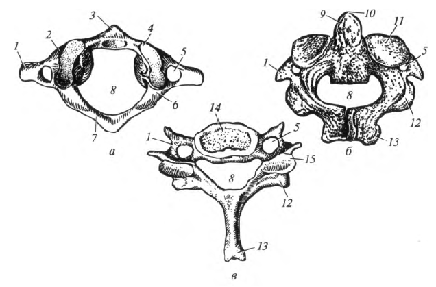
Fig. 4.7. Cervical vertebrae: a - atlas; b - axial; c - VII cervical; 1 - transverse process; 2 - lateral mass; 3 - front arc; 4 - upper articular surface; 5 - hole of the transverse process; 6 - furrow of the vertebral artery; 7 - a back arch; 8 - vertebral foramen; 9 - dentoid process (tooth); 10 - the tip of the tooth; 11 - upper articular surface; 12 - lower articular process; 13 - spinous process; 14 - the body of the vertebra; 15 - superior articular process
Transverse processes terminate in two tubercles - anterior and posterior. The anterior tubercle of the VI cervical vertebra is more developed than in others. It is called the “carotid tubercle,” since the carotid artery can be pressed against it during bleeding.
The spinous processes are short, directed slightly downward and bifurcated at the end. The spinous process of the VII cervical vertebra is longer, thickened at the end, so this vertebra is called "protruding" (its tip is well palpable under the skin).
The articular processes of the cervical vertebrae are short, located obliquely between the frontal and horizontal planes.
The shape of the first two cervical vertebrae was influenced by the immediate vicinity of the skull. With their participation, the head rotates, so they are called "rotational vertebrae."
I cervical vertebra - atlas, atlas (C I), has no body, is deprived of the spinous and articular processes. On the sides are lateral masses, the upper surfaces of which are articulated with the condyles of the occipital bone; the lower articular surfaces are slightly concave, articulate with the II cervical vertebra. The posterior arc of the atlas corresponds to the arc of a typical vertebra. On the upper surface of the arc posterior to the lateral mass there is a furrow of the vertebral artery. In place of the body, the atlas has an anterior arch, on which a platform is visible for connecting to the dentoid process (tooth) of the II cervical vertebra.
II cervical vertebra - axial, axis (С p), differs sharply from typical cervical vertebrae in that on the upper surface of his body there is a tooth-like process, or tooth, dens, which in development is a moving body of the atlas. When articulating the I and II cervical vertebrae, the tooth plays the role of an axis around which the atlas rotates with the skull. On the upper articular processes on the sides of the tooth there are articular surfaces for articulation with the lower articular fossa of the lateral masses of the atlas.
Thoracic vertebrae, vertebrae thoracicae (Th I -Th XII), significantly larger than the cervical. The height of the bodies of the thoracic vertebrae from I to XII is gradually increasing. Their transverse size also increases. The thoracic vertebrae are characterized by the presence of costal fossae located on the lateral surfaces of the body and transverse processes that serve to connect with the ribs (Fig. 4.8). The articular processes of the thoracic vertebrae stand in the frontal plane, the articular surface of the upper ones is turned back, the lower ones - forward. The transverse processes in front have an articular fossa for articulation with the tubercle of the rib. The spinous processes of the thoracic vertebrae are longer than those of the cervical vertebrae, tilted downward and tiled on top of each other.
Lumbar vertebrae, vertebrae lumbales (L I -L V), have a massive bean-shaped body. The height and width of the body gradually increase from I to V vertebra. The vertebral foramen is large compared to other vertebrae. The articular processes are well defined, their articular surfaces are located in the sagittal plane: in the upper processes they are directed medially, in the lower ones laterally. Transverse processes are located in the frontal plane, their ends are deflected posteriorly. Spinous processes of thin, flat, with thickened edges, located almost flush with the vertebral body.

Fig. 4.8. Thoracic vertebra (side view): 1 - upper articular process; 2 - upper vertebral notch; 3 - upper costal fossa; 4 - the body of the vertebra; 5 - lower costal fossa; 6 - lower vertebral notch; 7 - lower articular process; 8 - spinous process; 9 - transverse process; 10 - costal fossa
Sacrum, os sacrum, consists of five sacral vertebrae (S: - S v), which in an adult are fused into one bone. In the sacrum, the upper wide section is distinguished - the base, the lower - narrow - the apex; front (concave) - pelvic and posterior (convex) surfaces, as well as lateral (lateral) parts (Fig. 4.9).

Fig. 4.9. Sacrum and tailbone: a - front view; b - rear view; 7 - tailbone; 2 - the apex of the sacrum; 3 - transverse lines; 4 - front sacral openings; 5 - side part; 6 - the base of the sacrum; 7 - sacral tuberosity; 8- median sacral crest; 9 - lateral sacral crest; 10- intermediate sacral crest; 77 - the sacral canal; 12 - coccygeal horn; 13 - back sacral openings; 14 - ear surface
The base of the sacrum has upper articular processes that articulate with the lower articular processes of the V lumbar vertebra. The junction of the base of the sacrum with the body of this vertebra forms a ledge directed forward - the cape.
On the pelvic surface of the sacrum, horizontally oriented four transverse lines are visible - traces of the fusion of the bodies of the sacral vertebrae. At the ends of these lines, the front (pelvic) sacral openings open at the ends of the right and left - the exit points of the anterior branches of the sacral spinal nerves.
On the dorsal surface of the sacrum are the posterior (dorsal) sacral openings for the exit of the posterior branches of the sacral spinal nerves. Outside of the dorsal sacral openings are paired lateral parts on which to find
articular articular surfaces. The same surfaces of the pelvic bone are connected to them. Sacral tuberosity is located posterior to the articular surfaces.
When the sacral vertebrae grow together into a single bone, the vertebral openings form the sacral canal, ending at the bottom of the sacral fissure. Pelvic and dorsal sacral openings are connected to the sacral canal by intervertebral openings.
Coccyx, os coccygis, in an adult consists of 3 - 5 rudimentary vertebrae (Co: -Со ш). Only in the 1st coccygeal vertebra, in addition to the body, are the rudiments of the upper articular processes - the coccygeal horns, connected through ligaments to the sacral horns. The remaining vertebrae are rounded and small in size.
Ribs, costae, - bones connected in pairs with the thoracic vertebrae (12 pairs). Each rib has a posterior, longer, bone part and anterior, shorter, cartilaginous (costal cartilage). Seven pairs of upper ribs (I-VII) with cartilaginous parts connected to the sternum - true ribs. The cartilages of the VIII-X pairs of ribs are connected to the cartilage of the overlying ribs, forming false ribs; XI and XII pairs of ribs have short cartilaginous parts that end in the muscles of the abdominal wall - oscillating ribs (Fig. 4.10).
In the bone part of the rib, the head, neck and body are distinguished (Fig. 4.11). The head of the rib connects to the vertebral body. Behind the head, the posterior end of the rib narrows, forming the neck of the rib, which passes into the longest section - the body. Between the neck and body is a tubercle, which serves to articulate with the transverse process of the corresponding thoracic vertebra.
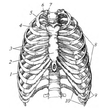
Fig. 4.10. Chest (front view): 1 - the xiphoid process; 2 - the body of the sternum; 3 - costal cartilage; 4 - the handle of the sternum; 5 - the body of the rib; 6 - neck of the rib; 7 - rib head; 8 - true ribs; 9 - false ribs; 10 - oscillating ribs

Fig. 4.11. I right (a) and II right (b) ribs: 1 - articular surface; 2 - rib head; 3 - neck of the rib; 4 - the articular surface of the tubercle of the ribs; 5 - angle of the rib; 6 - the body of the rib; 7 - a furrow of a subclavian vein; 8 - tubercle of the anterior scalene muscle; 9 - the furrow of the subclavian artery
The bodies of the II-XII ribs are curved anteriorly, have inner and outer surfaces, upper and lower edges. The rib bends toward the front, forming the angle of the rib. A groove of the rib for vessels and nerves runs along its lower edge.
Unlike the others, the ridge has upper and lower surfaces, medial and lateral edges. A tubercle is located on the upper surface for attaching the anterior scalene muscle. In front of the tubercle is the furrow of the subclavian vein, behind the furrow of the subclavian artery.
The sternum, sternum, is a flat bone located almost in the frontal plane. It consists of three parts: the upper - the sternum handle, the middle - the sternum body, the lower - the xiphoid process (see Fig. 4.10). Three notches are located on the upper edge of the sternum handle: in the middle - jugular, on the sides - paired clavicular (for articulation with clavicles); below the latter, on the lateral edge, there are recesses for cartilage of I-II ribs - rib cuts. The sternum body at the edges has cuttings for cartilage of the III - VII ribs. The xiphoid process is much narrower and thinner than the body, its shape is different: it is usually pointed downwards, sometimes has a through hole or bifurcated.
Spinal column
The back of the body is divided into two symmetrical halves by a longitudinal dorsal groove, which stretches from the neck to the pelvis. When the body is bent, the bony protrusions protrude one below the other in the dorsal sulcus. These are processes of the vertebrae - the bones from which the spinal column is formed; on the pelvis, the groove is smoothed and widened, forming a triangular wedge-shaped surface, under which the bone base is palpated; this triangle corresponds to the lower part of the spine - the sacrum.
The vertebral column is the main core of the entire skeleton, it connects the upper part of the skeleton with the lower one and is a flexible, unpaired bone formation consisting of separate bones - vertebrae, which are mostly movably connected, but in places, for example, on the sacrum, have grown together (Fig. . 8).
Fig. 8. The vertebral column (L); above - cervical, below - thoracic ECC below -belt *. the vertebrae and below - the sacrum with the tailbone:
1 - front surface 2 - back surface 3 - lateral, right surface. 4 - cervical bend. 5 - chest (dorsal) bend. 6 - lumbar bend
The vertebral column consists of 33-34 vertebrae. The structure of the vertebrae is generally the same, however, in different places of the trunk, the vertebrae have differences associated with functional load. In the neck, the spine is associated with neck movements and head movements, in the chest part, the spine is connected with the ribs, forming with them the rib cage; in the lumbar spine carries a greater load and has greater mobility than in the chest. This is where the mutual movements of the chest and pelvis occur.
In the sacral region, the spine participates in the formation of the pelvis and bears the entire heaviness of the upper part of the figure; the tailbone bends forward and is connected with the internal organs of the so-called small pelvis.
Thus, the vertebral column consists of 7 cervical, 12 thoracic, 5 lumbar, 5 sacral and 4-5 coccygeal vertebrae and, accordingly, this is divided into sections of the cervical, thoracic, lumbar, sacral and coccygeal. The length of the spine is 40% of the length of the whole body.
Due to the increasing downward loading, the size of individual vertebrae increases downward; the smallest vertebrae are cervical, the thoracic vertebrae increase downwards, the largest - lumbar.
The lumbar vertebrae rest on the sacrum, which is a triangular sphenoid bone facing down with the top of the clip; it is called the apex of the sacrum. The lower part of the spine follows - the coccyx, which forms, as it were, a continuation of the sacrum downward and consists of 4-5 bone little bodies fused together; coccyx with its lower narrow end pointing down, bent forward, tied

Fig. 8. (continued). The structure of the vertebra (chest), pitchfork from above (B); left view (B): I - vertebral body. 2 - intervertebral * opening, 3-transverse process. 4 - arc of the vertebra. 5 - spinous process. 6 - articular process. 7 - platform for articulation with a rib
with internal organs and to the formation of an external form has no relation.
As already mentioned, all individual vertebrae, with the exception of the two upper cervical vertebrae, have the same structure. In each vertebra there is a massive unpaired part facing forward, - body with an arch extending from it posteriorly and processes. From the arc backwards departs spinous process two transverse process go to the right and left, and up and down the right and left two articular processes. On the thoracic vertebrae there are also articular sites for articulation with ribs (Fig. 8). Between the vertebral bodies are cartilages fused with them, the so-called intervertebral discs; in addition, the vertebrae are connected by joints formed between the articular processes. The arc and body of each vertebra limit vertebral foramen thanks to which the vertebrae superimposed on each other together form located inside the spinal column spinal canal - container spinal cord.
Vertebrae are called from top to bottom: for example. II - cervical vertebra, III - thoracic, V - lumbar, etc. In a newborn, the spinal column is almost straight, but with age, under the influence of gravity, bends in the sagittal direction appear in it (Fig. 8). In the neck, the spine is bent forward, in the chest region - back, in the lumbar region - forward, the sacrum is bent back. Forward bend - lordosis bending backwards - kyphosis, peculiar to a normal body (of course, if they are not excessive); bending sideways - scoliosis arises due to improper development and is a deviation from the norm. These bends of the spine have a protective function - they, like springs, soften the tremors that occur during movements.
In general, the spine has great mobility. In it, movements of flexion, extension, tilts to the sides, rotation, circular motion, springy movements are possible.
The skeleton of the body is made up of the vertebral column and chest. Together with the medulla of the skull, they form the axial skeleton of the body, skeleton axiale.
The vertebral column is part of the axial skeleton and represents the most important supporting structure of the body, it supports the head, and limbs are attached to it. The movements of the body depend on the spinal column. The spinal column also performs a protective function in relation to the spinal cord, which is located in the spinal canal. The indicated functions are provided by the segmental structure of the spinal column, in which rigid and mobile-elastic elements alternate.
The length of the vertebral column in an adult male of average height (170 cm) is approximately 73 cm, with 13 cm in the cervical region, 30 cm in the thoracic, 18 cm in the lumbar region, and 12 cm in the sacrococcygeal. on average 3-5 cm shorter and 68-69 cm. The length of the spinal column is about 2/5 of the entire body length of an adult. In old age, the length of the spinal column decreases by about 5 cm or more due to an increase in the bends of the spinal column and a decrease in the thickness of the intervertebral discs.
The cervical, thoracic, lumbar, sacral and coccygeal parts are distinguished in the spinal column. The first three consist of divided vertebrae, interconnected by a complex system of joints. In the last two parts, complete or incomplete fusion of bone elements occurs, which is due to their predominantly supporting function.
The human vertebral column has a number of differences from the spine of animals. They are associated primarily with upright posture, in which the load on the vertebrae increases from top to bottom. In this direction, an increase in the vertebral bodies occurs. The sacrum is especially powerfully developed. The number of sacral vertebrae in humans, as in anthropoids, reaches 5-6, while in lower primates it usually does not exceed 3-4. A feature of the human sacrum is its large width, maximum in the upper section. On the other hand, in the process of evolution, there was a shortening of the cervical vertebrae and a decrease in the number of thoracic and lumbar vertebrae, as a result of which the total length of the human spinal column became smaller. Especially significant reduction of the vertebrae occurred in higher primates in the caudal region. The spinous processes of the vertebrae in humans are shorter and less massive than in anthropoids and primitive people, which is associated with a weaker development of the back muscles.
A characteristic feature of the human spinal column is its S-shape, due to the presence of four bends. Two of them are convex forward - this is the cervical and lumbar lordosis, and two are turned back - thoracic and sacral kyphosis. In mammals, the spine forms a weakly pronounced lordosis in the cervical part, and its trunk part has the appearance of an arc, which corresponds to the horizontal position of the body. The transformation of the spine in the direction of the formation of its bends begins already in monkeys. Anthropoids have a weak S-shaped curvature of the spinal column, lumbar lordosis is hardly outlined. The cervical and lumbar lordoses were also weakly expressed in paleoanthropes (Neanderthals); from this we can conclude that their body was not yet completely straightened.
Bends of the spinal column are outlined in the prenatal period. In a newborn, the spine has a slight dorsal curvature with mild lordosis and kyphosis. After birth, the shape of the spinal column changes due to the development of body statics. Cervical lordosis appears when the child begins to hold his head, its formation is associated with tension in the cervical and spinal muscles. Sitting increases kyphosis of the thoracic spine. Straightening the body, standing and walking cause the formation of lumbar lordosis. After birth, the curvature of the sacrum characteristic of a person intensifies, which the fetus has already had for 5 months. The final modeling of the cervical and thoracic bends occurs by the age of 7, and lumbar lordosis fully develops during puberty. The presence of bends increases the spring properties of the spinal column.
The severity of the bends of the spinal column is individually variable. In women, lumbar lordosis is more pronounced than in men. On living people, a weak positive relationship of the lengths of the lumbar and cervical lordosis is shown, negative - with the length of thoracic kyphosis.
There are several developmental options for lumbar lordosis based on a vertical lumbar index, that is, the percentage of the sum of the posterior heights of the bodies of the lumbar vertebrae to the sum of the anterior ones. His classification:
- curtorachia - up to 97.9,
- orthorachia - from 98 to 101.9,
- koylorakhiya - 102 and more.
Group variation of the lumbar index - from 95.8 to 106.8. Kurtorachidny type is typical for Europeans, some groups of American Indians, Masai, ortorachidny - Japanese.
The posture of a person depends on the shape of the spinal column. There are three forms of posture:
- normal
- with pronounced bends of the back,
- with smooth bends (the so-called "round back").
An increase in breast kyphosis leads to stoop. By the age of 50, the bends of the spine begin to smooth out. Some people in old age develop common kyphosis of the spinal column. The reason for these changes in posture is a flattening of the intervertebral discs, weakening of the ligamentous apparatus of the spine, and a decrease in the tone of the back extensor muscles. This is facilitated by a sedentary lifestyle, an improper mode of work and rest. Exercise allows you to maintain a long form of the spine and good posture. No wonder the military and athletes in old age maintains the correct posture of the body.
In addition to bends in the sagittal plane, the spinal column has a small frontal bend in the upper part of the thoracic region, which is called physiological, or aortic, ohm. Usually it is located at the level of III – V of the thoracic vertebrae, is convex to the right side, and is apparently associated with either the passage of the thoracic aorta at this level or the predominance of the right arm. Pronounced refers to pathology. It may be due to abnormalities in the development of the vertebrae.
Compounds of the vertebrae and the movement of the spinal column
The vertebrae are interconnected both continuously, through cartilage and other joints, and through joints. Intervertebral discs are located between the vertebral bodies. Each disk consists of a ring located on the periphery and a gelatinous core occupying the central part of the disk. There is often a small cavity inside the disc. The fibrous ring is built of plates, the arrangement of the fibers in which is similar to the orientation of the fibers in osteons. The gelatinous core consists of mucous tissue and can change its shape. When loading the spinal column, the internal pressure in the core rises, however, it cannot shrink. The intervertebral disc as a whole plays the role of a shock absorber during movements, thanks to it there is a uniform distribution of forces between the vertebrae. Through the intervertebral discs, up to 80% of the weight of the overlying parts of the body is transmitted.
The greatest height of individual discs in the cervical spine is 5-6 mm, in the chest - 3-4 mm, in the lumbar - 10-12 mm. The thickness of the disc changes in the anteroposterior direction: thus, between the thoracic vertebrae the disc is thinner in front, between the cervical and lumbar vertebrae, on the contrary, thinner in the back.
The ultimate compressive strength of the intervertebral discs is on average 69-137 kg / cm2,
whereas in vertebral bodies it is only 26 kg / cm2. Therefore, with excessive loads, such as, for example, in pilots during ejection, vertebral bodies are more often damaged than the discs connecting them.The ligamentous apparatus of the spinal column plays a large role in its stabilization. The rectified position of the body is maintained with little activity of the own muscles of the back. With maximum bending of the body, these muscles relax, and the entire load falls on the ligaments. Therefore, lifting weights in this position is dangerous for the ligaments and joints of the spine.
The movements of the spinal column are carried out due to intervertebral discs and arched joints. The latter are formed by the articular processes of neighboring vertebrae and belong to flat joints. The shape of the articular surfaces allows for combined sliding in various directions. A pair of arched joints together with the intervertebral disc forms a “segment of movement” of the spinal column. The movements in the segments are limited by ligaments, articular and spinous processes and other factors, therefore the range of movements in one segment is small. However, many segments take part in real movements, and their total mobility is very significant.
The following movements are possible in the spinal column under the action of skeletal muscles on it: flexion and extension, abduction and adduction (lateral flexion), twisting (rotation), and circular motion.
Flexion and extension occurs around the front axis. The amplitude of these movements is 170-245º. When bending the vertebral bodies lean forward, the spinous processes are removed from each other. The anterior longitudinal ligament relaxes, and the tension of the posterior longitudinal ligament, yellow ligaments, interspinous and supraspinatus ligaments inhibits this movement. During extension, the spinal column deviates backward, while all its ligaments are relaxed except for the anterior longitudinal one, which, when tensioned, inhibits the extension of the spinal column.
Lead and reduction are performed around the sagittal axis. The total range of motion is 165º. When abduction of the spinal column, the tension of the yellow ligaments, capsules of the arched joints and the transverse ligaments located on the opposite side, limit this movement.
The rotation of the spinal column has a total volume of up to 120º. During rotation, the gelatinous nucleus of the intervertebral discs plays the role of the articular head, and the tension rings of the intervertebral discs and yellow ligaments inhibit this movement.
The direction and amplitude of movements in different parts of the spinal column are not the same. The cervical vertebrae possess the greatest mobility. The Atlant and axial vertebra joints have a special device here. The atlantooccipital and atlantoaxial joints formed by them together form a complex combined multiaxial joint in which head movements in all directions occur. Atlas plays the role of a bone meniscus.
The joints of the atlas and the axial vertebra are supplemented by a highly differentiated ligamentous apparatus. It is necessary to highlight the transverse ligament of the atlas, which forms a synovial connection with the tooth of the axial vertebra and prevents its displacement back into the lumen of the spinal canal, where the spinal cord is located. Ligament ruptures and dislocations in the atlantoaxial joint pose a mortal danger due to possible damage to the spinal cord. Movements between the remaining cervical vertebrae occur around all three axes. The range of motion increases due to the relative thickness of the intervertebral discs. Bending forward is accompanied by a sliding of the vertebral bodies,
so that the overlying vertebra can bend over the edge of the underlying. In the cervical region, flexion by 70º, extension and rotation by 80º are possible.The mobility of the thoracic vertebrae is limited to thin intervertebral discs, the chest and the location of the articular and spinous processes. The amplitude of motion during bending is 35º, during extension - 50º, during rotation - 20º.
In the lumbar part of the spinal column, thick intervertebral discs allow flexion, extension and lateral flexion. Bending is possible at 60º, and extension at 45º. Rotation here is almost impossible due to the location of the articular processes in the sagittal plane. The most free movements between the lower lumbar vertebrae. Here is the center of most common body movements.
A characteristic feature of the spinal column is a combination of rotation with lateral flexion. These movements are possible to a greater extent in the upper parts of the spine and are very limited in its lower parts. In the thoracic part with lateral bending, the spinous processes turn towards the concavity of the spine, and in the lumbar, on the contrary, towards the bulge. The maximum lateral flexion occurs in the lumbar region and its connection with the thoracic spine. The combined rotation is expressed by turning the vertebral bodies in the direction of flexion.
The sacrococcygeal connection in young people, especially in women, also has some mobility. This is significant during childbirth, when under the pressure of the fetal head the coccyx deviates backward 1-2 cm and the exit from the pelvic cavity increases.
The range of motion of the spinal column decreases significantly with age. Signs of aging appear here earlier and are more pronounced than in other parts of the skeleton. These include degeneration of the intervertebral discs and articular cartilage. The intervertebral discs become more fibrous and, loosened, lose their elasticity and, as it were, are squeezed out of the vertebrae. Calcification of cartilage takes place, and in some cases, ossification appears in the center of the disc, which leads to the fusion of neighboring vertebrae. Following the disks, the vertebrae change. The vertebral bodies become porous, osteophytes form along their edges. The height of the vertebral bodies decreases, often they become wedge-shaped, which leads to a flattening of the lumbar lordosis. The width of the vertebrae in the frontal plane increases along the upper and lower edges; vertebrae take the form of "reel". Bone proliferation occurs along the edges of the articular surfaces of the vertebrae. One of the most frequent manifestations of spinal column aging is ossification of the anterior longitudinal ligament, which is well detected on radiographs.
Development and age features of the spinal column
The skeleton of the body undergoes in the embryonic development the blastema, cartilage and bone stages. Vertebrae and ribs have a distinct segmental arrangement, due to metamerism of the embryo body. An embryo forms segmental clusters of mesoderm on both sides of the chord called somites. The first pair of somites appears on the 16th day from fertilization, and at the end of the 6th week the embryo has 39 pairs of somites. From the total mass of the mesoderm, groups of cells stand out that form the rudiments of the axial skeleton, called sclerotomes.
Mesenchyma in sclerotomes is unevenly distributed; in the intervals between the somites there are clusters of cells representing the rudiments of the vertebral bodies, and at the level of the somites intervertebral discs are formed. Thus, the body of each vertebra develops due to two adjacent segments. Intersegmental artery enters the middle of the vertebral body. From the primary center,
located in the circumference of the chord, the mesenchyme extends dorsally to the neural tube, forming the rudiment of the arch and spinous process (the neural part of the vertebra), and laterally, giving rise to the transverse and costal processes.The blastema stage is replaced by cartilage. The cartilage first appears in the vertebral body, and then in the arch and costal processes: the latter are separated in the thoracic vertebrae, forming cartilaginous ribs, and in the cervical, lumbar and sacral vertebrae, separation of the costal processes does not occur. The cartilaginous vertebra is a single whole and is not subdivided into parts. In the early stages of development, the vertebral bodies of various departments have a similar shape.
Ossification of the vertebrae begins at the 2nd month of the embryonic period and occurs in the cranio-caudal direction. The first ossification points appear in the arches of the cervical vertebrae; at the 3rd month, the ossification points in the arches of the thoracic and lumbar vertebrae are laid. At the same time, the ossification of the rib ribs begins. In the vertebral bodies, ossification points appear earlier in the thoracic region (also at the beginning of the 3rd month). Ossification of the thoracic vertebrae and ribs can be considered as one of the signs of the beginning maturation of the functional respiratory system. At the 4th month, ossification points can be found in the bodies of the lumbar vertebrae, at the 5th month in the bodies of the cervical and sacral vertebrae. Ossification of the vertebral bodies occurs endochondrally, the formation of bone is preceded by the penetration of blood vessels into the cartilage. Later, a cortical plate of compact substance is formed by perichondral ossification. The chord is preserved as a gelatinous core. The ossification points in the bodies of typical vertebrae are laid symmetrically in each half of the body, but quickly merge with each other. In the arches of the vertebrae, the ossification points are paired.
In a newborn, a typical vertebra consists of three bone elements - the body and two halves of the arc, separated by layers of cartilage. On the upper and lower surfaces of the vertebral body there is also cartilage in the form of plates worn on the vertebra like canning lids. The intervertebral discs at this age are half the height of the vertebral bodies. Therefore, on radiographs of the spinal column of newborns between the vertebral bodies there are wide gaps that are occupied by the discs and the mentioned cartilage plates.
In view of these features, the spinal column of the fetus and newborn is elastic, but has low strength. Therefore, during childbirth, in cases of improper position of the fetus and with careless performance of obstetric procedures, injuries of the spinal column often occur, especially in its cervical part; this leads to damage to the spinal cord and the arteries supplying it and can cause a variety of nervous disorders and even death of the newborn.
After birth, the growth of the vertebrae in width occurs periostally, the growth in height is due to the formation of bones near the cartilaginous plates. By 3-4 months, the fusion of the half of the arches in the lower thoracic and upper lumbar vertebrae begins, from here the process spreads in both directions, and in the 2-3rd year of life, closure occurs almost throughout the entire spinal column. Following this, the spinous processes ossify. The fusion of vertebral bodies with arches occurs in the range of 3-6 years.
The ossification of some vertebrae differs from the given scheme. In Atlanta, the ossification points are laid in the anterior arch and lateral masses, the fusion of parts of the vertebra is completed by 10 years. The tooth of the axial vertebra has two independent ossification points that appear in the fetus for 4-5 months, a bit later an additional point is formed at the top of the tooth. Due to these points, the upper body of the axial vertebra is ossified; their synostosis with the vertebral body occurs in 4-5 years.
Pages: 1