TASTE AND SMELL
H. Altner, I. Beckh
13.1. Characterization of chemical sensations
The sensations of taste and smell are due to the selective and highly sensitive response of specialized sensory cells to the presence of molecules of certain compounds. More broadly, specific responses to chemicals such as hormones or neurotransmitters are common in many cells and tissues. However, taste and olfactory sensory cells act as exteroceptors; their responses provide important information about external stimuli, processed by special areas of the brain, which are responsible for the corresponding sensations. Other chemoreceptor cells serve as interoceptors that determine, for example, the level of CO 2 (Section 21.6).
Taste and smell can be characterized and distinguished based on morphological and physiological criteria. The differences between these two types of sensations are most obvious when comparing the types (qualities) of stimuli adequate for them (Table 13.1). Other characteristics such as sensitivity to stimuli or the physical state the latter, although in general they are not the same, but can also overlap.
Compared to other senses, taste and smell have a significantly higher adaptability (cf. fig. 8.5). With prolonged exposure to the stimulus, excitation in the afferent pathways is noticeably weakened, and accordingly perception is weakened; for example, already very quickly in an environment, even with a strong smell, we cease to feel it. Equally characteristic of chemical sensations is a high sensitivity to certain stimuli. At the same time, the range of distinguishable stimulation intensities is relatively small (1: 500), and the discrimination threshold is high. Stevens exponentψ = k(φ - φо)aequal to 0.4–0.6 for smell and about 1 for gustatory stimuli (cf. Fig. 8.14).
Primary processes and chemical specificity .
The first event during the stimulation of chemoreceptors is, according to modern concepts, a chemical interaction based on the weak binding of an adequate molecule with receptor protein. Proteins with enzymatic properties were isolated from the taste organs, substrate
the specificity and kinetic features of which are the same as those of the receptors themselves. Subsequent events leading to an electrical reaction cell membrane, are unknown. Each receptor cell reacts highly selectively to a specific group of substances. The slightest changes in the structure of a molecule can change the way it is perceived or make it an inadequate stimulus. The stimulatory efficacy of a compound is probably most significantly dependent on its size (e.g. chain length) and internal distribution. electric charges(i.e., the location of the functional groups). However, the fact that in many cases the molecules of substances that differ greatly in chemical structure, cause the same olfactory sensations, has not yet been explained. For example, the three substances below, despite their structural differences, have the same musky odor (see. Beets's).
It was suggested that chemoreceptors contain receptor centers, specific for certain groups of substances. This point of view is confirmed by cases of partial anosmia, that is, insensitivity to the smell of some very close chemical compounds... Similarly, one can interpret the selective effect of certain drugs on the taste organ. Applying potassium hymnemate (a substance isolated from an Indian plantGymnema silvestre) leads to the loss of only the perception of sweetness - sugar makes the mouth feel gritty. The protein found in the fruits of the West African plantSynsepalium dulcificum, changes the sour taste to the sweet one, so that the lemon is perceived as an orange. Kurihara v ). Cocaine on the tongue causes a consistent loss of all four types of taste sensations: bitter, sweet, salty, and finally sour.
Table 13.1.Classification and characteristics of chemical senses
|
Taste |
Smell |
|
|
Receptors |
Secondary sensory cells |
Primary sensory cells; endings |
|
Localization of receptors |
Language |
V, IX and X cranial nerves, Nose and pharynx |
|
Afferent cranial nerves |
VII, IX |
I, V, IX, X |
|
Levels of synaptic switching in the central nervous system |
1 medulla oblongata 2.Ventral thalamus 3. Cortex (postcentral gyrus) C link with the hypothalamus |
1. Olfactory bulb 2.The forebrain (prepiriform cortex) Connections with the limbic system and hypothalamus |
|
Adequate incentives |
Molecules organic and inorganic substances, mostly non-volatile. Source of stimuli - near or in direct contact with receptors |
Almost exclusively molecules of organic volatile substances in the gas phase, dissolving only near the receptor. The source of incentives is usually removed |
|
Number of qualitatively distinguishable stimuli |
Few (4 main) |
A very large (thousands) many indistinct qualities |
|
Absolute sensitivity |
Comparatively low (At least 10 16 molecules in 1 ml of solution) |
Very high for some substances (10 7 molecules, in animals - up to 10 2 -10 3 molecules in 1 ml of air) |
|
Biological characteristic |
Contact feeling. Used to assess food quality, regulate food intake and digestion (salivary reflexes) |
Distant feeling. Used for hygienic assessment of the environment and food; in animals, for the search for food, communication and in sexual behavior. Includes a strong emotional component |
In adults sensory taste cells located on the surface of the tongue. Together with supporting cells, they form groups of 40-60 in the epithelium of its papillae. elements - taste buds(fig.13.1). The large papillae surrounded by a roller at the base of the tongue contain up to 200 taste buds each; in smaller mushroom and leaf-like papillae on its anterior and lateral surfaces, there are only a few of them. In total, an adult has several thousand taste buds. Glands, located between the papillae, secrete the liquid that washes the taste buds. The distal parts of the receptor (sensory) cells sensitive to stimulation form microvilli, going into a common chamber, which communicates with the external environment through a pore on the surface of the papilla (Fig. 13.1). Stimulating molecules diffuse through this pore and reach the taste cells (receptors).
Like other secondary sensory cells, gustatory cells generate receptor potential upon stimulation. This excitement is synaptically transmitted afferent fibers
Rice. 13.1.Scheme of the taste bud immersed in the papilla of the tongue; shows the basal, sensory, supporting cells and afferent fibers of the corresponding cranial nerve
cranial nerves, which conduct it to the brain in the form of impulses. This process involves: drum string-branch of the facial nerve(Vii) innervating the anterior and lateral parts of the tongue, and glossopharyngeal nerve(IX), innervating its posterior part (Fig. 13.2). Branching out, each afferent fiber receives signals from the receptors of different taste buds.

Rice. 13.2.Human language scheme. Its innervation is highlighted by coloring by various cranial nerves; the contours show the distribution areas of different types of papillae (1-mushroom, 2-surrounded by a roller, 3-leaf-shaped). Localization of zones of perception of certain taste qualities is shown by icons
Replacedtaste cells very quickly; the lifespan of each of them is only about 10 days, after which a new receptor is formed from the basal cell. It establishes a connection with afferent fibers in such a way that their specificity does not change. The mechanism providing such interaction is still not clear (see. Oakley's).
Cell-to-fiber reactions . Single taste cell in most cases it reacts to substances of different taste, depolarizing or hyperpolarizing by them (Fig. 13.3). The amplitude of the receptor potential increases with the concentration of the stimulating substance. The type and amplitude of the response is also influenced by the environment (Figure 13.4).
The generator potential causes the appropriate level of excitation of the afferent fibers, forming a reaction called the "gustatory profile" (Fig. 13.5). Their impulse depends on the reaction of the receptors as follows: depolarization of the latter has an exciting effect, hyperpolarization has an inhibitory effect.
Many fibers of the IX pair of cranial nerves react particularly strongly to substances with a bitter taste. Fibers of the VII pair are more excited by the action of salty, sweet and sour: some of them react more strongly to sugar than to salt, others to salt than to sugar, and so on. These taste-specific features

Rice. 13.3.Intracellular recordings of the receptor potentials of two taste cells (a, b) of the rat tongue. Stimuli: 0.5 mol / L NaCl; 0.02 mol / l quinine hydrochloric acid; 0.01 mol / l HCI and 0.5 mol / L sucrose. The duration of each stimulus is shown horizontal line(on Sato, Beidler in with changes)

Rice. 13.4. Influence of the environment on the shape and amplitude of intracellular recordings of the receptor potential of a single taste cell of the rat tongue stimulated by 0.02 mol / L quinine hydrochloric acid. Environment: a - 41.4 mmol / l NaCl; b - distilled water (by Sato, Beidler in with changes)

Rice. 13.5.Responses of two single fibers of a rat drum string to various substances: 0.1 mol / l NaCl;
0.5 mol / L sucrose; 0.01 N. HCI; 0.02 mol / l quinine hydrochloric acid (as modified)
different afferent groups provide information about the taste, those. the form of a stimulating molecule, and the general level of excitement of a certain population of them is about the intensity of the stimulus, that is, about the concentration of a given substance.
Central neurons. Flavoring fibers VII and IX pairs of cranial nerves terminate within or in the immediate vicinity of single path nuclei ( nucleus solitarius ) medulla oblongata. This nucleus is connected through the medial loop (medial lemniscus) to thalamus in its ventral posteromedial nucleus. Axons of third-order neurons pass through the inner capsule and end in postcentral gyruscerebral cortex. V As a result of information processing at the listed levels, the number of neurons with highly specific gustatory sensitivity increases. A number of cortical cells respond only to substances with one flavor quality. The location of such neurons indicates a high degree of spatial organization of the sense of taste. Other cells in these centers respond not only to taste, but also to temperature and mechanical stimulation of the tongue.
Taste qualities . Man distinguishes four main taste qualities: sweet, sour, bitter and salty, which are well characterized by their typical substances (Table 13.2). The sweet taste is associated mainly with natural carbohydrates such as sucrose and glucose; sodium chloride — salty; other salts, for example KCI are perceived as salty and bitter at the same time. Such mixed feelings are characteristic of many natural gustatory stimuli and correspond to the nature of their components. For example, orange is sweet and sour, and grapefruit
Table 13.2.Substances with a characteristic taste and the effectiveness of their effect on humans ( Pfaffmann's)
|
Quality |
Substance |
Perception threshold, mol / l |
|
Bitter |
Quinine sulfate |
0,000008 |
|
Nicotine |
0,000016 |
|
|
Sour |
Hydrochloric acid |
0,0009 |
|
Lemon acid |
0,0023 |
|
|
Sweet |
Sucrose |
0,01 |
|
Glucose |
0,08 |
|
|
Saccharin |
0,000023 |
|
|
Salty |
NaCI |
0,01 |
|
CaCI 2 |
0,01 |
sour-sweet-bitter. Acids taste sour; many plant alkaloids are bitter.
On the surface of the tongue can be distinguished zones of specific sensitivity. Bitter taste is perceived mainly basis language; other tastes affect it lateral surfaces and tip, moreover, these zones overlap (Fig. 13.2).
Between chemical properties substance and its taste there is no one-to-one correlation. For example, not only sugars, but also lead salts are sweet, and artificial sugar substitutes such as saccharin have the sweetest taste. Moreover, perceived quality substance depends on its concentration. Table salt in low concentration appears sweet and becomes purely salty only when it is increased. Sensitivity to bitter substances is significantly higher. Since they are often poisonous, this feature warns us against danger, even if their concentration in water or food is very low. Strong bitter irritants easily cause vomiting or urge to her.Emotional components taste sensations vary widely depending on the state of the organism. For example, a person who is deficient in salt finds the taste to be acceptable, even if the concentration in the food is so high that normal person refuses to eat.
The mouthfeel is obviously very similar. in all mammals. Behavioral experiments have shown that different animals have the same taste characteristics as humans. However, registration of the activity of individual nerve fibers also revealed some abilities that are absent in humans. For example, cats have "Water fibers" either responding only to water irritation, or exhibiting a taste profile that includes water as an effective stimulus (cf. Sato's).
Biological significance . Biological role taste sensation lies not only in checking the edibility of food(see above); they also affect the digestion process. Connections with vegetative efferents allow gustatory sensations to influence the secretion of the digestive glands, and not only on its intensity, but also on the composition, depending, for example, on whether sweet or salty substances predominate in food.
With agethe ability to discriminate taste is reduced. The consumption of biologically active substances such as caffeine and heavy smoking lead to the same.
The surface of the nasal mucosa is enlarged due to the nasal concha-ridges protruding from the sides into the lumen of the nasal cavity. Olfactory area, containing most of the sensory cells,

Rice. 13.6.Diagram of human nasopharyngeal cavities (sagittal incision). The olfactory area is limited by the upper and middle shells. Areas innervated by the trigeminal (V), glossopharyngeal (IX) and vagus (X) nerves are shown
bounded here by the superior turbinate, although in the middle there are also small islands of the olfactory epithelium (Fig. 13.6).
The olfactory receptor is a primary bipolar sensory cell, from which two processes extend: from above - a dendrite carrying cilia, from the base of the axon. Cilia, the internal structure of which is different than that of ordinary kinocilia, are immersed in the layer of mucus covering the olfactory epithelium and are not able to actively move. The odoriferous substances carried by the inhaled air come into contact with their membrane — the most likely site of interaction between the stimulating molecule and the receptor. Axons heading to the olfactory bulb are combined into bundles ( fila olfactoria ). In addition, free endings are found throughout the nasal mucosa. trigeminal nerve, and some of them also react to smells. In the pharynx, olfactory stimuli are able to excite fibers of the glossopharyngeal and vagus nerves (Figure 13.6). The layer of mucus covering the olfactory epithelium protects it from drying out and is constantly renewed due to secretion and redistribution by kinocilia.
Odor molecules enter to receptors (sensory cells) periodically: during inhalation through the nostrils and, to a lesser extent, from the oral cavity, diffusing through the choanae. Thus, when we eat, we have mixed sensations, which combine the taste and smell of food.
Rice. 13.7.Simultaneous recording of an electro-olfactogram (Red line) and the action potentials of a single receptor of the frog olfactory epithelium upon stimulation with nitrobenzene. Stimulus duration (black line)–1 s ( Gesteland's)
Sniffing — a characteristic behavior of many mammals — significantly increases the flow of air to the olfactory mucosa and hence the concentration of stimulating molecules in it.
In total, a person has about 10 7 receptors in the olfactory region with an area of about 10 cm 2. In other vertebrates there are much more of them (in the German shepherd, for example, 2.2-10 8). Olfactory cells, like taste cells, are constantly being replaced and because of this, apparently, not all function at the same time.
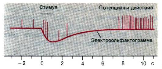
Electrodes placed on the olfactory epithelium of vertebrates, under the action of smell, register slow potentials of a complex shape with an amplitude of several millivolts. These electroolfactograms(EOG, Fig.13.7, see. Ottoson c), like electroretinograms (ERG), reflect the total activity of many cells, therefore, they do not provide information on the properties of individual receptors. Record Activity single receptor in the olfactory mucosa of vertebrates it was possible only by chance (Fig. 13.7). It has been shown that the spontaneous activity of these cells is very low (several pulses per second), and each of them reacts to a variety of substances. As with the flavor profile, you can build range of responses single olfactory receptor (see. Gesfeland c).
A person is able to distinguish the smell of thousands of different substances. Olfactory sensations can be classified on the basis of a certain similarity, identifying certain types, or quality, smell. However, this is much more difficult to do than in the case of gustatory sensations. The uncertainty of the categories is obvious also in the fact that the classifications proposed by different authors are not the same. Correlation between chemical structure and the quality of the odor is even less than in the case of gustatory irritants. Tab. 13.3 shows that classes of odors are usually named after their natural
Table 13.3.Distinctive characteristics of odor classes ( Amoor, Skramlik)
|
Odor class |
Known Typical Substances |
Similarity to smell |
"Standard" source |
|
Floral |
Geraniol |
Roses |
d –1 – β – phenylethylmethylcarbinol |
|
Ethereal |
Benzyl acetate |
Pears |
1,2-dichloroethane |
|
Musky |
Muscon |
Musk |
3-methylcyclopentadecane-1-one |
|
Camphor |
Cineol, camphor |
Eucalyptus |
1,8-cineole |
|
Putrefactive |
Hydrogen sulfide |
Rotten eggs |
Dimethyl sulphide |
|
Pungent |
Formic acid, acetic acid |
Vinegar |
Formic acid |
sources or typical substances; each category can also be characterized by a “standard” source.
The neurophysiological basis for classifying odors into one class or another has not yet been discovered. The point of view, according to which the groups uniting substances similar in smell, are somewhat different from each other, is confirmed by cases of partial impairment of the sense of smell (partial anosmia). With such defects (at least some of them of a genetic nature), the threshold for perception of certain olfactory stimuli increases. At the same time, it often changes for several substances, which, as a rule, belong to the same class of odors. Experimental data used to classify odors can be obtained by analyzing cross adaptation. It lies in the fact that when the prolonged action of any smell causes an increase in the threshold of its perception, the sensitivity to the smell of some other substances also decreases (Fig. 13.8). By studying quantitatively such mutual changes in thresholds, it is possible to construct a scheme of cross-adaptive relationships (Figure 13.9). However, this is not enough for an unambiguous and detailed classification of a variety of odorous substances according to the sensations they cause.
When interpreting the features of the human sense of smell, it should be borne in mind that endings are also sensitive to odorous substances trigeminal nerve in the nasal mucosa, as well glossopharyngeal and vagus nerve in the throat. All of them are involved in the formation of the olfactory sensation (Fig. 13.6). Their role, which has nothing to do with the olfactory nerve, is also preserved when the function of the olfactory epithelium is impaired due to, for example, infection (influenza), tumors (and related operations on the brain) or traumatic brain injury. In such cases, combined by the term hyposmia, the threshold of perception is significantly higher than normal, but the ability to distinguish odors decreases only slightly. With pituitary hypogonadism (Kalman's syndrome), the sense of smell is provided exclusively by these cranial nerves, since in this congenital disease, aplasia of the olfactory bulbs occurs. Harmful temperature and chemical influences can cause reversible or irreversible acute or chronic hyposmia or anosmia, depending on the nature and method of exposure. Finally, the sensitivity to odors decreases with age.
Human sense of smell is very sensitive, although it is known that in some animals this apparatus is even more perfect. Table 13.4 shows the concentration of two odoriferous substances, sufficient to cause the corresponding sensations in a person. Under the action of very small amounts of them, the sensation that occurs is nonspecific; only after exceeding a certain threshold level, the smell is not only detected, but also recognized. For example, skatole in low concentrations smells quite acceptable; at higher ones, repulsive. Thus, it is necessary to distinguish detection threshold and recognition threshold smell.
Such thresholds, determined by the responses of the subjects or the behavioral reactions of animals, do not allow to establish single sensory cell sensitivity(receptor). However, knowing the spatial extent of the human olfactory organ and the number of receptors in its composition, it is possible to calculate their sensitivity. Such calculations show that a single sensory cell is depolarized and generates an action potential in response to one, at most a few molecules of an odorous substance. Of course, a behavioral response requires the activation of a significant number of receptors, i.e. exceeding a certain critical level of the signal-to-noise ratio in afferent fibers.
Coding.So far, the coding of olfactory stimuli by receptors can only be described in a first approximation. First, an individual sensory cell is capable of responding to many different odorous substances. Secondly,

Rice. 13.8.An increase in the intensity of sensation with an increase in the intensity of stimulation (with propanol) without adaptation (black straight line) and after adaptation to pentanol (red curve with black triangles) ( Cain, Engen in with changes)

Rice. 13.9.Cross-adaptive relationships of seven odorous substances: 1-citral, 2-cyclopentanone, 3-benzyl acetate, 4-safrole, 5-m-xylene, 6-methyl salicylate, 7-butyl acetate. Reciprocal interactions are usually unequal. The degree of increase in the perception threshold is indicated as follows: black lines are very large; red solid lines are large; dashed red lines — moderate; red dotted line - weak(software with changes)
Table 13.4.The detection threshold for odors of butyric acid and butyl mercaptan ( Neuhaus, Stuiver)
|
Substance g |
The number of molecules in 1 ml of air |
Concentration substances near source of stimulus, mol / l |
|
Butyric acid |
2,4–10 9 |
10 –10 |
|
Butyl mercaptan |
10 7 |
2,7– 10 –12 |
different olfactory receptors (as well as gustatory receptors) have overlapping response profiles. Thus, each odoriferous substance specifically excites an entire population of sensory cells; in this case, the concentration of the substance is reflected in general level excitement.
Central processing of olfactory information
Olfactory bulb . Histologically, the olfactory bulb is subdivided into several layers, characterized by cells of a specific shape, from which branches of a certain type depart with characteristic connections between them. The main features of information processing are as follows: significant convergence sensory cells on the mitral; expressed braking mechanisms and efferent control input impulse. In the glomerular (glomerular) layer, the axons of approximately 1000 receptors end on the primary dendrites of one mitral cell(fig.13.10). These dendrites also form reciprocal dendrodendritic synapses with periglomerular cells. Mitral – periglomerular contacts – excitatory, opposite in direction – inhibitory. The axons of the periglomerular cells end on the dendrites of the mitral cells of the neighboring glomerulus. Such an organization allows modulating the local dendritic response, providing auto-inhibition and inhibition of surrounding cells. Cells-grains also form dendrodendritic synapses with mitral cells (in this case, with their secondary deidritis) and thereby affect their generation of impulses. Inputs on mitral cells are also inhibitory, i.e. reciprocal contacts are involved in auto-inhibition. Finally, grain cells form synapses with mitral cell collaterals, as well as with efferent (bulbopetal) axons of various origins. Some of these centrifugal fibers come from the contralateral bulb through the anterior commissure (commissure).
The peculiarity of the inhibition caused by the cells-grains deprived of axons is that, in contrast to the typical inhibition according to Renshaw, they can be activated partially, that is, with a spatial gradient. This

Rice. 13.10.Diagram of neural connections in the olfactory bulb. In the glomeruli (glomeruli), the axons of the olfactory receptors end in primary dendrites ( D 1) mitral cells. Periglomerular cells and granule cells form reciprocal synapses with primary and secondary ( D 2) mitral cell dendrites. K-collaterals. The direction of synaptic transmission is shown by arrows: exciting influences - black, brake –Red(software with generalizations and changes)
the picture of very complex interactions is quite comparable to that known in the retina, although information processing is based on a different principle of cellular organization. Everything described above is just a rough diagram of the events taking place in the olfactory bulb. In addition to mitral, secondary neurons also include a variety of bundle cells, which differ in their projections and mediators.
Central connections . Axons mitral cells form lateral olfactory tract, bound for prepiriform bark and piriform share. Synapses with higher-order neurons provide communication with hippocampus and, through the amygdala, with vegetative nuclei hypothalamus. Neurons that respond to olfactory stimuli are also found in reticular formation midbrain and in orbitofrontal cortex.
The effect of smell on other functional systems . The direct connection with the limbic system (see Section 16.6) explains the pronounced emotional component olfactory sensations. Smells can cause pleasure or disgust (hedonic components), influencing accordingly the affective state of the body. In addition, the importance of olfactory stimuli in regulation of sexual behavior, although the results of experiments on animals, especially experiments on the blockade of smell in rodents, cannot be directly transferred to humans. It has also been shown in animals that responses from neurons in the olfactory tract can be altered by injection of testosterone. Thus, sex hormones also affect their arousal.
Functional disorders . In addition to hyposmia and anosmia, there is a misperception of smell. (iarosmia) and olfactory sensation in the absence of odoriferous substances (olfactory hallucinations). The causes of these disorders are varied. For example, they can occur with allergic rhinitis and head injuries. Olfactory hallucinations of an unpleasant nature (cacosmia) are observed mainly in schizophrenia.
Tutorials and Guides
1. Beidler LM.(Ed.). Chemical Senses. Part 1. Olfaction. Part 2. Taste. Handbook of Sensory Physiology, Vol. IV, Berlin – Heidelberg – New York, Springer, 1971.
2. PfaffD.(Ed.). Taste. Olfaction and Central Nervous System. New York, Rockfeller University Press, 1985.
Original articles and reviews
3. Breipohl W.(Ed.). Olfaction and Endocrine Regulation, London, IRL Press, 1982.
4. Denton D. A., Coghlan J. P.(Eds.). Olfaction and Taste, Vol. V, New York, Academic Press, 1975.
5. Hayushi T. (Ed.). Olfaction and Taste, Vol. II, Oxford – London New York – Paris, Pergamon Press, 1967.
6. Kare M. R., Mailer 0.(Eds.). The Chemical Senses and Nutrition, New York – San Francisco London, Academic Press, 1977.
7. Koster E. Adaptation and Cross Adaptation in Olfaction, Rotterdam, Bronder, 1971.
8. Le Magnen J., Mac Lead P.(Eds.). Olfaction and Taste., Vol. VI, London – Washington DC, IRL Press, 1977.
9. Norris D. M.(Ed.). Perception of Behavioral Chemicals, Amsterdam – New York – Oxford, Elsevier / North Holland, 1981.
10. Pfaffman WITH. (Ed.). Olfaction and Taste, Vol. Ill, New York, Rockfeller University Press, 1969.
11. Sato T. Receptor potential in rat taste cells. In: Autrum H., Ottoson D., Perl E. R., Schmidt R. F., Shimazu H., Willis W.D.(Eds.). Progress in Sensory Physiology, Vol. 6, p. 1–37, Berlin – Heidelberg – New York – Tokyo, Springer, 1986.
12. Schneider D.(Ed.). Olfaction and Taste, Vol. IV, Stuttgart, Wiss, Verlagsges, 1972.
13. Shepherd G. M. Synaptic organization of the mammalian olfactory bulb. Physiol. Rev. 52, 864, (1972).
14. Van der Starre H.(Ed.). Olfaction and Taste. Vol. VII, London – Washington DC, IRL Press, 1980.
15. Zotterman Y.(Ed.). Olfaction and Taste. Vol. I. Oxford – London – New York Paris, Pergamon Press, 1963.
16. Chemical Senses. London. IRL Press (Published in regular installments).
Why is the same dish perceived differently by different people? For example, the soup seems to you wonderful in its original form, and your soul mate wants to pepper or salt it. In the first case, we are talking about the category of people "supermasters", on whose tongue there are many papillae and the taste seems complete. As a rule, "supermasters" prefer soft food instead of spicy, and they like to add cream to coffee. The category of people with a low density of papillae - "subteysters" - love spicy food that "burns" the oral cavity. Although taste sensitivity is influenced not only by the papillae.
It is known that the human brain distinguishes five tastes - sour, salty, bitter, sweet and umami (rich exotic taste of oriental food). However, the set of chemicals that trigger these signals differs from one to another. different people... Experts point out that humans possess between 20 and 40 genes responsible for detectors of bitter taste.
The different perception of bitter is most likely a consequence of evolutionary pressure in different parts of the planet. The most poisonous plants are endowed with a bitter taste, and nomadic tribes who came into contact with different types of plants, over time, received different receptors.
People in countries where malaria is common most often have a gene that makes them less sensitive to certain bitter substances, especially those containing cyanide. Researchers believe that small amounts of cyanide can neutralize malaria insects without poisoning humans. By the way, people have a natural aversion to bitterness and some odors, which is why the beer, beloved by many, is rarely liked by anyone at the first try.
If you want to understand who you are - "supermaster" or "subteyster" - put food with blue dye on your tongue. This blue dye does not stick to the taste buds on the tongue. If there is little blue left on the language, you are a "supermaster"; if the whole language is blue, you are a "subteyster."
Table of contents of the subject "Vestibular sensory system. Taste. Gustatory sensitivity... Olfactory sensory system. Odor (smells). Odor classification. ":1. Vestibular sensory system. Function of the vestibular system. Vestibular apparatus. Bone labyrinth. Membranous labyrinth. Otoliths.
2. Hair cells. Properties of receptor cells of the vestibular apparatus. Stereocilia. Kinocilius.
3. Otolith apparatus. Otolith organ. Adequate stimuli for the receptors of the otolith organs.
4. Semicircular canals. Adequate stimuli for the receptors of the semicircular canals.
5. The central part of the vestibular system. Vestibular nuclei. Kinetoses.
6. Taste. Gustatory sensitivity. Gustatory sensory system. Taste reception. Taste time.
8. Central department of the gustatory system. Pathways of taste sensitivity. Cores of taste.
9. Taste perception. Olfactory sensory system. Macrosmatics. Microsmatics.
10. Smell (s). Odor classification. Stereochemical theory of odors.
Membrane of microvilli of taste cells contains specific sites (receptors) designed to bind dissolved in liquid medium oral cavity chemical molecules. There are four types of taste sensations, or four taste modalities: sweet, sour, salty, and bitter. A strict relationship between chemical nature of the substance and no taste: for example, not only sugars have a sweet taste, but also some inorganic compounds(salts of lead, beryllium), and the sweetest substance is saccharin, which cannot be absorbed by the body. Most taste cells are polymodal, that is, they can respond to stimuli from all four taste modalities.
Joining specific receptors molecules with a sweet taste activates the system of secondary messengers adenylate cyclase - cyclic adenosine monophosphate, which close the membrane channels of potassium ions, and therefore the membrane of the receptor cell is depolarized. Substances with a bitter taste activate one of two systems of secondary mediators: 1) phospholipase C - inositol-3-phosphate, which leads to the release of calcium ions from the intracellular depot with subsequent release of the mediator from the receptor cell; 2) the specific G-protein gastducin, which regulates the intracellular concentration of cAMP, which controls the cation channels of the membrane and this determines the emergence of the receptor potential. The action on the receptors of molecules with a salty taste is accompanied by the opening of gated sodium channels and depolarization of the taste cell. Substances with a sour taste close membrane channels for potassium ions, which leads to depolarization of the receptor cell.
The magnitude of the receptor potential depends on taste and concentration chemical acting on the cell. The emergence of a receptor potential leads to the release of a neurotransmitter by the taste cell, acting through the synapse on the afferent fiber of the primary sensory neuron, in which the frequency of action potentials increases 40-50 ms after the onset of the stimulus. Nerve impulses arising in afferent fibers are conducted to the nuclei of single bundles of the medulla oblongata. With an increase in the concentration of the active substance, the total number of responsive sensory fibers increases due to the involvement of high-threshold afferents in the transmission of information from receptors.
Gustatory sensitivity
Gustatory thresholds are detected by alternately applying solutions of substances with different taste qualities to the surface of the tongue (Table 17.4). The absolute threshold of sensitivity is the appearance of a certain taste sensation that differs from the taste of distilled water. Taste the same substance can be perceived differently depending on its concentration in solution; for example, at low concentrations of sodium chloride, it tastes sweet, and at higher concentrations, salty. The maximum ability to distinguish between the concentration of solutions of the same substance and, accordingly, the lowest differential threshold of taste sensitivity is characteristic for the middle range of concentrations, and at high concentrations of the substance, the differential threshold increases.
Table 17.4. Absolute thresholds for the perception of substances with a characteristic taste
Absolute gustatory thresholds individually differ, but the overwhelming majority of people have the lowest detection threshold for substances with a bitter taste. This feature of perception arose in evolution, it contributes to the rejection of eating substances of bitter taste, to which the alkaloids of many poisonous plants belong. Taste thresholds differ in the same person depending on his need for certain substances, they increase due to prolonged use of substances with characteristic taste(for example, sweets or pickles) or smoking, alcohol consumption, burning drinks. Different areas of the tongue differ in taste sensitivity to various substances, which is due to the peculiarities of the distribution of taste buds. The tip of the tongue is more sensitive to sweets than other areas, the sides of the tongue to sour and salty, and the root of the tongue to bitter. Taste sensations in most cases are multimodal and are based not only on the selective chemical sensitivity of taste receptor cells, but also on food irritation. thermoreceptors and mechanoreceptors of the oral cavity, as well as the action of volatile food components on olfactory receptors.
Determines the formation of gustatory sensations, is a reflexogenic zone. With the help of the taste analyzer, various qualities of taste sensations, the strength of sensations, which depends not only on the strength of irritation, but also on the functional state of the body, are assessed.
Structural and functional characteristics of the taste analyzer.
Peripheral department... Taste receptors (taste cells with microvilli) are secondary receptors; they are an element of taste buds, which also include supporting and basal cells. The taste buds contain cells containing serotonin and cells that form histamine. These and other substances play a role in shaping the sense of taste. Individual taste buds are polymodal formations, as they can perceive different kinds taste irritants. Taste buds in the form of separate inclusions are located on the back of the pharynx, soft palate, tonsils, larynx, epiglottis and are also part of the taste buds of the tongue as an organ of taste.
The peripheral section of the taste analyzer is represented by taste bulbs, which are located mainly in the papillae of the tongue. Taste cells are dotted at their end with microvilli, which are also called taste hairs. They come to the surface of the tongue through the gustatory pores.
The taste cage has big number synapses that form fibers drum string and glossopharyngeal nerve. The fibers of the tympanic string (a branch of the lingual nerve) fit all the mushroom papillae, and the fibers of the glossopharyngeal nerve - to the grooved and leaf-shaped. The cortical end of the gustatory analyzer is located in the hippocampus, parahippocampal gyrus, and in the lower part of the posterior central gyrus.
Taste cells continually divide and die continually. The replacement of cells located in the front of the tongue, where they lie more superficially, occurs especially quickly. Replacement of taste bud cells is accompanied by the formation of new synaptic structures
Conductor department... Inside the taste bud are nerve fibers that form receptor-afferent synapses. Taste buds of different areas of the oral cavity receive nerve fibers from different nerves: taste buds of the anterior two-thirds of the tongue - from the drum string, which is part of the facial nerve; the kidneys of the posterior third of the tongue, as well as the soft and hard palate, tonsils - from the glossoglossal nerve; taste buds, located in the region of the pharynx, epiglottis and larynx, from the superior peglottic nerve, which is part of the vagus nerve.
These nerve fibers are the peripheral processes of bipolar neurons located in the corresponding sensory ganglia, representing the first neuron of the conduction part of the taste analyzer. The central processes of these cells are part of a single bundle of the medulla oblongata, the nuclei of which represent the second neuron. From here, the nerve fibers in the medial loop approach the optic hillock (third neuron).
Central department. The processes of thalamic neurons go to the cerebral cortex (fourth neuron). The central, or cortical, department of the gustatory analyzer is localized in the lower part of the somatosensory cortex in the region of the tongue. Most of of neurons in this area is multimodal, that is, it reacts not only to gustatory, but also to temperature, mechanical and nociceptive stimuli. It is characteristic of the gustatory sensory system that each taste bud has not only afferent, but also efferent nerve fibers that approach taste cells from the central nervous system, which ensures that the taste analyzer is included in the integral activity of the body.
The mechanism of taste perception... For a gustatory sensation to arise, the irritant must be in a dissolved state. A sweet or bitter flavoring substance that dissolves into molecules in saliva penetrates into the pores of taste buds, interacts with the glycocalyx and is adsorbed on the cell membrane of the microvilli, into which “sweet-feeling” or “bitter-sensitive” receptor proteins are embedded. When exposed to salty or sour flavors, the concentration of electrolytes around the taste cell changes. In all cases, the permeability of the cell membrane of microvilli increases, sodium ions move into the cell, depolarization of the membrane occurs and the formation of a receptor potential, which spreads both along the membrane and along the microtubular system of the taste cell to its base. At this time, a mediator (acetylcholine, serotonin, and, possibly, hormone-like substances of a protein nature) is formed in the taste cell, which in the receptor-afferent synapse leads to the emergence of a generator potential, and then an action potential in the extrasynaptic parts of the afferent nerve fiber.
Perception of gustatory stimuli. Microvilli of taste cells are formations that directly perceive the taste stimulus. Membrane potential taste cells range from -30 to -50 mV. Under the action of gustatory stimuli, a receptor potential of 15 to 40 mV arises. It is a depolarization of the surface of the taste cell, which is the cause of the appearance of a generator potential in the fibers of the tympanic string and glossopharyngeal nerve, which, when a critical level is reached, turns into propagating impulses. From the receptor cell, excitation is transmitted through the synapse to the nerve fiber with the help of acetylcholine. Some substances, such as CaCl 2, quinine, heavy metal salts, cause not primary depolarization, but primary hyperpolarization. Its occurrence is associated with the implementation of negative rejected reactions. In this case, no propagating impulses arise.
Sensitivity of receptors to different types taste irritations.
Different taste cells have different sensitivity to different taste substances, which are divided into four groups: sour, salty, sweet, bitter. Each cell always responds to more than one flavoring agent, sometimes even to all four, but it has the greatest sensitivity to one of them. Accordingly, depending on the location of cells with a particularly high sensitivity to a particular taste stimulus, different parts of the tongue also have different sensitivity.
It has been established that the tip of the tongue and its anterior third are most sensitive to sweets, where mushroom papillae are located, the lateral surfaces to sour and salty (leaf-shaped papillae), and the root of the tongue to bitter (grooved papillae, or taste buds surrounded by a shaft).
Gustatory cells are characterized by fluctuations in the threshold of irritation and different in different conditions the nature of the response to the same stimuli. Their excitability depends on constant influences on each other, as well as on the state of the receptors of the digestive tract, olfactory organs, etc. Normally, there is a certain "tuning" of taste receptors in accordance with the state of the body, in particular with the state of satiety.
Send your good work in the knowledge base is simple. Use the form below
Students, graduate students, young scientists who use the knowledge base in their studies and work will be very grateful to you.
Posted on http://www.allbest.ru/
Department of Physiology
Physiology of taste
Introduction
1. Morphology of the organs of taste; subjective physiology of taste. Orientation and structure of taste buds
2. Central connections
3. Basic taste sensations
4. Intensity of sensations
5. Objective physiology of taste
6. Primary process
7. The role of gustatory sensitivity
Literature
Introduction
Man and animal continuously receive information about the endless variety of changes that occur in the external and internal environment. This is due to the presence of specialized structures in the body, which are called analyzers (sensory systems).
Analyzers are understood as a set of formations that ensure the perception of stimulus energy, its transformation into specific processes of excitation, the conduction of this excitation into the structures of the central nervous system and to the cells of the cortex, analysis and synthesis of this excitation by specific zones of the cortex, followed by the formation of sensation.
The concept of analyzers was introduced into physiology by I.P. Pavlov in connection with the doctrine of higher nervous activity... Each analyzer consists of three sections:
The peripheral or receptor section, which perceives the energy of the stimulus and transforms it into a specific process of excitation.
The conduction section, represented by afferent nerves and subcortical centers, carries out the transmission of the arisen excitement to the cerebral cortex.
The central or cortical part of the analyzer, represented by the corresponding areas of the cerebral cortex, where the higher analysis and synthesis of excitations and the formation of the corresponding sensation are carried out.
The role of analyzers in the formation of adaptive reactions is extremely large and diverse. According to the concept of the functional system of P.K. Anokhin, the formation of any adaptive reaction is carried out in several stages. Analyzers are directly involved in the formation of all stages of the functional system. They are suppliers of afferent parcels of a certain modality and various functional purposes, and the same afferentation can be situational, starting, reverse and indicative, depending on the stage of formation of adaptive activity.
taste physiology analyzer organ
1. Morphology of the organs of taste; subjective physiology of taste.Orientation and structure of taste buds
The tongue in humans is covered with a mucous membrane, the folds of which in many places form small protuberances in the form of pegs, called papillae.
These three types are distributed in different ways. Only mushroom papillae are scattered over the entire surface. Grooved papillae, of which a person has only 7-12, from above have the appearance of round formations 1-3 mm in diameter; they are located in a limited area across the dorsum of the tongue at its root. The third type, leaf-shaped papillae, form tightly spaced folds along the posterior edges of the tongue. They are well developed in children, but much less pronounced and less numerous in adults.
The filiform papillae that occupy the rest of the tongue are not shown in Fig. 1 because they lack taste buds. The name "kidney" refers to the shape of these organs (Fig. 2). Their position on the papillae varies; in the case of grooved and leaf-shaped papillae, many taste buds are embedded in the side walls, but there are none at the apex. In mushroom papillae, taste buds are limited by the surface of the mushroom cap, which can be up to 1 mm in diameter.
The individual taste bud is about 70 µm in height and about 40 µm in diameter. In total, a person has about 2000 taste buds, of which about half are on grooved papillae. Each taste bud contains 40-60 individual cells.
V connective tissue under the grooved and leaf-shaped papillae, serous glands are immersed, the ducts of which open into depressions at the base of the papilla, their secret serves to wash off food particles and microorganisms. In addition, it lowers the concentration of the stimulant near the taste buds.
Inside the taste buds, three types of cells are distinguished: sensory, supporting, and basal (Fig. 2). Water-soluble substances that fall on the surface of the tongue diffuse through the pore into the liquid-filled space above the taste bud; here they come into contact with the membranes of the microvilli, which form the outer ends of the sensory cells. Taste receptors are secondary sensory cells without axons that conduct impulses in a central direction. Their responses are transmitted by afferent fibers that form synapses near the bases of sensory cells. In fig. 2 shows only two fibers, but in reality about 50 fibers enter and branch into each taste bud.
The lifespan of sensory cells in taste buds is short; there is a continuous change. On average, one sensory cell is replaced by a new one within 10 days. The change of cells can be traced by marking their nuclei with 3H-thymidine and determining the number of labeled nuclei preserved after some time. Lost sensory cells are replaced by new ones, which are formed from basal cells. With this change, synapses between afferent fibers and old cells should be interrupted and new synapses should arise. In connection with such a restructuring, many interesting questions, especially considering the fact that sensory cells differ in their sensitivity to different stimuli. Thus, a change in sensory cells can lead to a change in the "taste profile" - a characteristic form of responses in afferent fibers, which will be discussed in the next section.
2. Central connections
Afferent fibers conducting responses from accumulations of taste buds are distributed along two cranial nerves - the facial (VII) and glossopharyngeal (IX). This division usually corresponds to the regions of the tongue that are supplied with these fibers. So, the fibers from the grooved and leaf-shaped papillae go mainly as part of the glossopharyngeal nerve, and the fibers from the mushroom papillae in the front of the tongue enter the drum string (chorda tympani), a branch of the facial nerve. Children have additional taste organs in the epithelium of the soft palate and the posterior wall of the pharynx to the larynx; they are innervated mainly by the vagus nerve (X).
In the brain, taste fibers on each side combine to form a solitary tract. It ends in the medulla oblongata, in the nucleus of the solitary tract, where afferent fibers form synapses with second-order neurons. The axons of these neurons go to the ventral thalamus as part of the medial lemniscus. The third set of neurons connects this area with the cerebral cortex. The gustatory zones of the cortex are located in the lateral part of the postyntral gyrus.
3. Basic taste sensations
Under normal conditions, such as eating, the oral mucosa is exposed to complex stimuli that include several modalities. Due to the fact that the oral cavity communicates with the nasal cavity, odoriferous substances can reach the olfactory receptors in the nose and cause other sensations. In addition, the mucous membrane of the mouth and tongue contains thermoreceptors, mechanoreceptors, and pain fibers, which are also stimulated. What is commonly referred to as "taste" is actually a multimodal sensation in which sensations of smell, warmth or cold, pressure and possibly even pain are superimposed on the actual taste sensations.
There are four distinct basic taste sensations: sweet, sour, salty, and bitter.
The detection thresholds for different qualities are at different concentrations. The threshold concentration of quinine sulfate (8 μmol / L, or 0.006 g / L) is a good example that bitter tasting substances are found at very low concentrations. The detection threshold for saccharin is 23 μmol / L (0.0055 g / L), for grape sugar - 0.08 mol / L, and for cane sugar - 0.01 mol / L (14.41 and 3.42 g, respectively) / l). These data are representative and show that the thresholds for mono- and disaccharides are significantly higher than for synthetic sweets. The thresholds for acetic acid (0.18 mol / L, or 0.108 g / L) and table salt (0.01 mol / L, or 0.585 g / L) illustrate the general rule that the thresholds for acidic and salty are about the same order of magnitude. as for the above saccharides. Acid thresholds roughly reflect the degree of acid dissociation. Comparison of the thresholds for grape and cane sugar suggests that the grape sugar solution must be more concentrated than the cane sugar solution in order for them to be equally sweet. Experimental verification of solutions of different above-threshold concentrations corresponds to this difference.
But the usefulness of such accurate cut-off data is limited because, for most substances, the cut-off levels are subject to significant individual variability. It would be more reasonable to talk about the range of threshold values
4. Intensity of sensations
A simple comparison of different solutions shows that the intensity of the taste sensation depends on the concentration of the substance. When determining the thresholds, it was found that the effect of diluting a solution of a stimulating substance can be compensated by stimulating a larger surface of the tongue, i.e. a greater number of receptors This is probably due to spatial summation. In the threshold region, there is an input relationship between the concentration and the duration of the stimulus. In addition, it should be remembered that the sense of taste is subject to a certain adaptation - with prolonged action of the stimulus, the intensity of the sensation decreases. Another factor is the secretion of the serous glands, which dilutes the substance acting on the taste buds and thus influences the intensity of the sensation.
Testing of a number of dilutions of saline solutions in the near-threshold region in many cases shows that sensation can change its quality depending on concentration. Table salt solutions 0.02-0.03 mol / l have a sweet taste, and at a concentration of 0.04 mol / l or more, they are salty. This shift in quality can probably be understood from the fact that taste fibers have a wide range of sensitivities within each quality.
Different areas of the tongue in humans vary in sensitivity to four basic qualities. The tip of the tongue is especially sensitive to sweet substances; the middle portions of the edges respond best to sour stimuli. Saline stimuli are most effective in the region of the tongue edge, which partially overlaps the first two. Bitter substances act most strongly on receptors near the root of the tongue, in the region of the grooved papillae. Therefore, damage to the glossopharyngeal nerve reduces the ability to detect bitterness, and after blockade of conduction in the facial nerve, only they are found.
5. Objective physiology of taste
The ability to discriminate tastes depends on the specificity of receptor molecules in the membranes of sensory cells. Microelectrodes can be used to record the activity of both individual sensory cells and afferent fibers. Such records show that neither the receptors themselves nor the fibers going to the central nervous system produce qualitatively specific responses; incentives in more than one category are generally effective. It is obvious that each fiber responds to stimuli of several categories, but when considering different gradations of sensitivity, differences are revealed. In other words, stimulation with a solution of a substance at a certain concentration activates various fibers in varying degrees... The arousal pattern typical of each individual fiber in response to a range of substances is called the taste profile. The fibers closest to qualitative specificity are those that respond to sugar solutions by increasing the discharge frequency. Comparative studies have shown that such relatively specific fibers are especially characteristic of monkeys.
Registration of the activity of individual sensory cells showed that they have a gradual relative specificity. The responses of the fibers emanating from these cells, in this respect, reflect the responses of the cells. But afferent fibers branch out in taste buds, so that each fiber receives excitation from many sensory cells, which, presumably, differ in the degree of specificity. In addition, it was found that sensory cells in different papillae form synapses with collaterals from one afferent fiber. This means that taste fibers receive inputs from sensory cells distributed over large areas of the tongue. These areas are called receptive fields. The situation with receptive fields is complicated by the fact that individual sensory cells can receive innervation from several different fibers.
The gradual relative specificity of taste fibers is created by 1) the gradual relative specificity of sensory cells and 2) the branching of taste fibers that creates receptive fields. The frequency of impulses in a single afferent fiber therefore varies with both the quality of the stimulus and its concentration. Of course, the extent to which the stimulated area covers the fiber's receptive field is also an important factor. The obvious takeaway with regard to stimulus coding is that the activity of a single fiber cannot provide unambiguous information about quality or concentration. Only a comparison of the level of arousal in several fibers can reveal characteristic distributions (patterns) of activity that say something about the quality of the stimulus. Provided the quality is known, the frequency of pulsing in each individual fiber can serve as a measure of the concentration of the stimulant. Distinctive features substances are therefore encoded in such a way that a complex but characteristic pattern of excitation is generated by the simultaneous but different responses of many neurons.
6. Primary process
The condition for excitation of the taste bud is the interaction between the molecules of the stimulating substance and specially differentiated points in the membrane of the sensory cell, where the receptor molecules lie. This interaction is called the primary process; it is believed to begin with the adsorption of the stimulus molecule. It is believed that when this happens, the receptor-probably protein-molecule changes its structure. This conformational change in the receptor molecule leads in turn to a local change in the permeability of the cell membrane. This cellular "amplifying mechanism" could be the reason for the generation of the receptor potential.
Evidence for the existence of specific receptor molecules includes the observation that certain plant substances and drugs, such as cocaine and hymnic acid (obtained from the Indian plant Gymnema sylvestre), selectively block some taste sensations. Obviously, this acid binds to receptor molecules for sweet substances, since its application makes such substances tasteless. The primary process in the membranes of taste sensory cells has not yet been fully explained, but according to the working hypothesis, it is similar to the process in cholinergic synapses, where special molecules change permeability at specific points in the membrane.
7. The Role of Gustatory Sensitivity
The taste buds on the tongue respond to stimuli located in the mouth. In other words, gustatory sensitivity in all vertebrates is involved in orientation at close range. At the same time, in fish, the sense of taste can also serve as orientation at a distant distance. In water, flavoring substances are transported by diffusion and convection from very distant sources to the flavoring bulbs, which can be dispersed over the entire surface of the fish's body.
In addition to its role in orientation at close range, a person's sense of taste has an important function, triggering a number of reflexes. For example, washing the tongue with secretions from the serous glands is controlled by a reflex, which is under the influence of taste buds. The secretion of saliva is also triggered by reflexive stimulation of the taste buds. Even the composition of saliva varies depending on the nature of the stimuli acting on the sensory cells, and gustatory stimuli also affect the secretion of gastric juice. Finally, it has been shown that vomiting is caused by the participation of gustatory sensitivity.
Literature
1. Batuev A.S., Kulikov G.L. Introduction to the physiology of sensory systems. - M .: graduate School, 1983.-263 p.
2. Lectures on physiology of central nervous system: Tutorial... Faculty of Biology and Chemistry of UdSU, Pronichev I.V. - Powered by swift.engine.edu, 2003 .-- 162 p.
3. Shulgovskiy V. V. Fundamentals of neurophysiology: textbook for university students. - M .: Aspect Press, 2000. p. 277.
4. Shulgovskiy VV Physiology of higher nervous activity with the basics of neurobiology: Textbook for student biol. specialties of universities. - M .: Publishing Center "Academy", 2003. - 464 p.
Posted on Allbest.ru
Similar documents
Inhomogeneous structure of the organ of taste. About 2,000 taste buds are found in the tissue of the tongue, palate, epiglottis, and upper esophagus. Most of them are located in the mucous membrane of the taste bud. Nerve fibers and the formation of taste buds.
abstract, added 03/02/2009
General physiology of sensory systems. Somatosensory, gustatory and olfactory analyzers. Determination of touch points. Determination of spatial thresholds of tactile reception and localization of pain receptors. Determination of taste sensations and thresholds.
manual, added 02/07/2013
general characteristics organism of a dog, features of its anatomy and physiology, functions of individual organs. Description of the main systems of the body: the system of bones, muscular, cutaneous and nervous. Features of the organs of sight, taste, hearing, touch and smell.
abstract, added 11/09/2010
Anatomy and physiology of the cardiovascular system. Veins, distribution and blood flow, regulation of blood circulation. Blood pressure, blood vessels, arteries. Determination of the indicator of the state of posture and flat feet in students. The organ of taste, types of papillae.
term paper added 12/25/2014
Study of the features of the development technology, types (syrup, injections, inhalations, granules, ointment, gel) and the composition of dosage forms for children. Characteristics of methods for determining the taste of drugs, numerical indices and organoleptic assessment of corrigens.
abstract, added 01/27/2010
The vestibular cochlear organ (organ of hearing and balance): the structure and interaction of elements, functions in the life of the human body. Propagation of sound in the organ of hearing. The location of the organ of smell and taste, the patterns of their functioning.
presentation added on 08/27/2013
The structure and physiology of the heart, its main functions. Description of the scheme and mechanism of blood circulation. Phases of the cardiac cycle, electrical activity of myocardial cells and parameters of central hemodynamics. The concept and features of the process of innervation of the heart.
presentation added on 01/12/2014
Normal physiology. Pathological physiology. Chronological table... Classification by groups and subgroups. Chemical structure, mechanism of action. Sources of origin, etc. The mechanism of biological activity of drugs in this group.
term paper, added 07/03/2008
Study of the anatomy and physiology of ENT organs as distant analyzers. Anatomy of the ear, nose, pharynx, larynx. Physiology of the nose and paranasal sinuses, auditory and vestibular analyzer. Respiratory, protective and voice-forming functions of the larynx.
abstract, added 01/29/2010
The structure of the diencephalon. The role of the liver and pancreas in digestion. Inhibition of the central nervous system. Anatomy and physiology of the autonomic nervous system, its age features... Composition of blood and physicochemical properties of plasma.