A rather large proportion of the mass of dry mycelium of fungi is their cell wall, namely, oh 5 to 15%. Its composition varies greatly and is often very specific for certain taxonomic groups of fungi. This can be seen from examples of its composition in representatives of yeast, chytrid and mucor fungi - zygomycetes, given in table. 1.3 (Aronson, 1965).
* (Amount of ash without phosphates.)
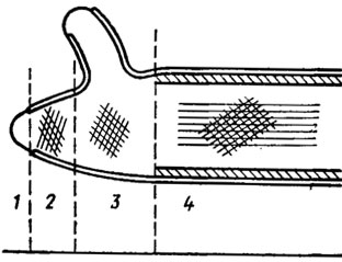
Structurally, the shells of the fungi are based on a two-phase system in which there are microfibrils included in the amorphous mass of the matrix. According to electron microscopy, it consists of at least two layers with differently directed orientation of the fibrils. The inner layer is usually oriented along the main axis of the cell, the outer ones are at an angle to it (Fig. 1.1). In yeast, the shell is usually multilayered, with mannan localized in the outer thick layer and glucan in the inner thin layer. In aquatic mushrooms, for example, in Allomyces, the membrane forms false septa - pseudosepts resembling wheel spokes. In marsupials and basidiomycetes, real septa are observed - septa. In marsupials, the septa between the cells usually have one simple pore, on both sides of which during its formation can be seen by a pair of osmophilic bodies of Voronin. In basidiomycetes, these pores are often very complex, equipped with caps — parentosomes (Fig. 1.2). Such pores were found in fruiting bodies and rhizomorphs of a number of higher basidiomycetes (Moore, 1965; Burnett, 1968). However, to date, it remains not entirely clear whether this difference in the structure of the septal pores of asco- and basidiomycetes can be attributed to their taxonomic affiliation or to the haploid and homocaryotic structure of the marsupial genome and to the di- and heterocaryotic nature of basidiomycetes. There are no such studies yet, but their significance for evolutionary and taxonomic constructions in the kingdom of mushrooms is very fundamental.

A lot of new things were added to the study of septal pores of fungi studies at the electron microscope level (Flegler et al., 1976; Kamaletdinova, Vasiliev, 1982). First, it became known that dolipores of basidiomycetes are structures that apparently hermetically isolate mycelial cells from each other until the formation of fruiting bodies by them (Flegler et al., 1976). This isolation is carried out by soluble proteolytic enzymes and at the same time osmophilic (containing proteins and lipids) bilateral caps, which disappear at the time of formation of fruiting bodies. Their disappearance is accompanied by perforations of the parentosomes and the communication between hyphal cells is restored.
Recent observations of septa in the fruit bodies of discomycetes (for example, Peziza badia - Kamaletdinova, Vasiliev, 1982) have shown that similar uncoupling hyphal cells of the structure also exist in the class of marsupials. Voronin bodies involved in flask-like invaginations of the cell membrane take part in their formation, the osmophilic contents of which (Voronin body) are released near the septum and are located at the septum opening, gradually penetrating into it, creating its closure device. The further penetration of Voronin's body into the overlying hymenial layer is apparently hindered by a special perforated structure lying above the septum in the mother cell of the future bag, which is completely isolated from the penetration of submicroscopic organelles from the subhymenial layer. Similar insulating structures are also observed in the paraphyses formed in the fruiting bodies.
Similar structures are also found in the sporulating mycelium of imperfect fungi containing Voronin bodies, for example, in Arthrobotrys conoides. In the penicillin producer, deuteromycete Penicillium chrysogenum, a structure was found in the septum that exactly corresponded to that found by Kamaletdinova and Vasiliev in discomycetes Peziza badia (Kurilowich et al., 1980).
The skeletal bases of the shells of the fungi are composed of crystalline organized polysaccharides: cellulose, chitin, chitosan, mannan, glucans and others. All of them have a linear structure with β-1,4 bonds of the starting components — hexose monomers, amino and acetaminohexoses. According to the results of microchemical testing (ruthenium red color), it was previously believed that pectin was present in the cell walls of fungi. However, the results of chemical analysis did not confirm the galacturonic acid monomer included in the pectin structure (Aronson, 1965).
Chitin and chitosan for most fungi are very characteristic in the composition of their shells as nitrogen-containing polymers. Moreover, the chitin of fungi is very similar to the chitin of insects and crustaceans, which was confirmed by the X-ray diffraction pattern. However, nitrogen in the chitin of fungi is less than in animals, and methylpentose, called mycetosis, was found among its components. Chitin in fungi can be detected microchemically by the method of Van Wisseling, using partial alkaline deacetylation and subsequent reaction to chitosan and X-ray diffraction. It was not found only in oomycetes, such as saprolegni and peronospore mushrooms. It was previously believed that chitin is absent in yeast, but it is found in cell walls - septa of saccharomycetes (Kulaev, 1975).
It has now been established that chitin can be found in Chitrydiales, Monoblepharidales, Protomycetales, Hyphochyiridiales, in all Endomycetales, Blastocladiales, Mucorales, Entomophthorales, in all marsupials and basidiomycetes, and Fungi imperfecti (deuteromes) derived from them. The exception is Oomycetes, in which polysaccharides contain cellulose in the membrane, which is completely absent in representatives of yeast fungi.
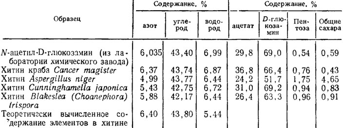
In recent years, in connection with the identification of the possibility of the practical use of fungal chitin for the synthesis of polymers, quite a lot of studies have appeared with data on a more subtle study of it in comparison with crustacean chitin (Table 1.4). Aspergillus niger has the least amount of acetate and D-glucosamine due to the higher content of pentoses and glucose in the composition of β-glucan and two α-glucans of the fungus shell. Analysis of diffraction patterns showed an identical crystalline structure of chitin of fungi and crabs with a slightly higher ordering in the latter. In addition, the chitin of fungi, in contrast to the lamellar structure of crab chitin, had a fibrous structure (Feofilova et al., 1980).
The chitin substitute for some mushroom fungi, chitosan, detected by weak acid hydrolysis by reaction to chitosan sulfate, was found, in addition to Mucor rouxii, also in Phycomyces blakesleeanus. The number of acetyl groups in it turned out to be different and varied in M. rouxii to zero. Among other aminopolysaccharides, a galactosamine polymer with a free amino group capable of binding phosphates with a structure such as chitosan was isolated from cell walls of Neurospora crassa, N. sitophitla, A. niger, and Botrytls drierea (Aronson, 1965). A number of fungi have polymers from amino sugars associated with mannan, glucan and proteins.
Cellulose in the cell walls of fungi usually does not occur simultaneously with chitin. An exception is one Rhizidiomyces from the order of hyphochitridium fungi in which they were found simultaneously. Cellulose was found in fungi from oomycetes of the orders Acrasiales, Lagenidiales, Saprolegniales, Leptomitales, Peronosporales (Aronson, 1965).
Among fungi that live in the aquatic environment, cellulose is usually found only in groups with bicameral zoospores. Blastocladiales and Monoblepharidales, which have single-flagged zoospores, do not. The exception that Rhizidiomyces is from the order of Hyphochytridiales, which has both chitin and cellulose and is considered to be a transitional form between the chitin Chytridiales and Blastocladiales and cellulose-containing oomycetes, is understandable. This one-flagellate flagellum has a feathery zoospore flagellum rather than a scab similar to Blastocladiales and Monoblepharidales. It is curious that the structure of the villi of the feathery flagella of the oomycetes resembles the structure of the flagella of bacteria, while the scab-like ones are completely similar to flagellate flagella.
Cellulose in fungi is easily detected by microchemistry using Speicher’s reagent or a reagent consisting of a solution of iodine in potassium iodide with the addition of a 70% solution of sulfuric acid.
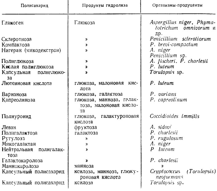
Glucans in mushrooms are very plentiful and differ from cellulose in their structure. Their monomers are also glucose. However, the most studied yeast glucans have less crystalline structures than cellulose. The crystallinity of glucans increases due to the formation of hydroglucans during their treatment with sulfuric acid. Similar glucans, in association with proteins, have been found in yeast and Penicilliunt notatum (Aronson, 1965). Another glucan, which is part of the shells of fungi, is callose, similar to that found in the sieve tubes of higher plants and strongly colored by basic dyes, i.e., having an acidic nature, in contrast to cellulose, has β-1,3-glucosidic bonds. A similar glucan stained with basic dyes was found in Sclerofinia. Glucans are also found in Aspergillus fischeri, Allomyces macrogynus, Neurospora crassa. In fungi, heteropolysaccharides composed of monomers of various sugars are also found, especially frequent in the genus Penicillium. In yeast-like forms pathogenic for animals, like Coccidioides and Cryptococcus, similar, but acidic polysaccharides are part of the capsule surrounding their cells. Examples of glucans and other products of the polymerization of monosugars and sugar acids that are part of the cell membranes and spare substances of fungi are given in table. 1.5.
Mannans, polysaccharides composed of mannose monomers. found especially abundantly in yeast, and they are frequent in species of yeast that live on the surface of the cambial standing under the bark of trees. Among these forms is the Hansenula living under the bark of conifers; in the capsule surrounding these yeast cells, the polysaccharide is present in the form of phosphomannan. This hydrophilic and mucous polysaccharide sticks together with yeast to the bristles covering the body of bark beetles, and in this way the yeast is transferred with their help from one tree to another (Wickerham, Barton, 1961). Mannans have not yet been found in hyphoblasting fungi, but mannose is found in hydrolysates of their cell walls.
In fungi, polysaccharides containing galactose, 6-deoxypentose, methylpentose, and most often fructose are also found, especially in mucorids. In the membranes of Penicillium chrysogenum cells, pentoses of 6-deoxypentosis, ramnose and xylose were also found from the polypore Polysiictus sanguineus.
Polysaccharide-protein complexes were found in yeast, for example, in Candida albicans, the causative agent of thrush in infants. The mannan-protein complex was found in Saccharomyces.
The lipids in fungi vary greatly quantitatively depending on environmental conditions and the age of the culture. Sometimes their number reaches 35-36% of the dry matter mass of cells. In yeast, more than 3% of lipids are found in their shells. They were also found in the shells of mukorovyh fungi, for example, in Musorouxii and Phycomyces, in the sporangiophores of which about 25% of lipids from their dry weight were found. They are probably found there in cuticle type formations (Aronson, 1965).
Pigments are also included in noticeable amounts in the composition of the cell walls of fungi. Pigments, especially the black pigment, often melanin, which is often localized as a special layer, are very often found in the cell walls of the mycelium or in the spore membranes of many fungi. Such a melanin layer is present in the shells of ascospores of Neurospora tetrasperma (Aronson, 1965).
Melanin is absent in fungi with low polyphenol oxidase activity, which takes part in its biosynthesis, and with a predominance of active dehydrogenases in the metabolism. Such fungi, which include representatives of the genera Fusarium, Trichothecium, Arthrobotris, Cephalosporium, and many others, are most often characterized by pink or orange coloration of sporulating structures, which depends on the abundance of carotenoids, which assume the role of a light shield and antioxidant, which belongs to dark-colored fungi melanin. The association of the presence of carotenoids with a high level of dehydrogenase activity is explained by the fact that the optimal conditions of the redox regime for the action of dehydrogenases coincide with the conditions optimal for the biosynthesis of carotenoids and other products of the terpenoid shunt. Carotenoid pigments are usually no longer part of the cell walls of fungi, but are localized either in the cell membrane or in drops of lipids dispersed in the cytoplasm. In some cases, completely special pigments are found in fungi, such as ommochromes, insect eye pigments, in the tinder fungus Pycnoporus (Polyporus) cinnabarinus (Shivrina, 1965) or phycobilins in Russula emetica and R. paludosa russula species, which are part of the photosynthesis system in blue-green and red algae.
Cell wall. It is a multilayer shell of 9 ... 10 layers of different electron density. A system of microfibrils embedded in an amorphous matrix forms the skeleton of a cell. Fibrils, depending on the species, may consist of cellulose, glucon and chitin. Other polysaccharides, proteins, pigments, lipids serve as cementitious substances that form chemical bonds with the microfibrillar part of the cell wall. The presence of such complexes provides selective permeability for some substances and blockade of others.
Supporting microfibrils of the cell wall and its matrix differ in the mechanism of formation and biosynthesis. The formation of fibrils and matrix occurs non-synchronously, the fibrillar skeleton of the wall is primarily regenerated. The biosynthesis of these two parts of the cell wall is carried out with the participation of enzymes.
The process of cell wall formation occurs in two ways: a new material can either penetrate into the wall polarized or evenly overlap over its entire surface. In the first case, the formation of cylindrical cells occurs, in the second - spherical.
The cell wall serves as a protective device and protects the fungal cell from exposure to various environmental factors, for example, the osmotic barrier, which determines the selective permeability of various substances. It gives shape to the vegetative cells of hyphae and the reproductive organs. Enzymes are localized on the surface of the cell wall and cytoplasmic membrane, which transform polymers that are not absorbed by the cell (insoluble in water).
As a result of lysis, the cell wall of fungi can collapse under the influence of enzymes secreted by other cells and formed in the cell of the fungus itself.
The main components of the cell wall of fungi are chitin, glucans, protein and fats. Nitrous and nitrogen-free polysaccharides with fatty substances form soluble and insoluble complexes. The basis of the cell wall is 4 ... 6 monosugars, the ratio of which varies slightly among different fungi. The polysaccharide fractions include glucosamine, mannose, glucose, xylose, etc. It should be emphasized that the composition of the cell membrane of various cells of the same fungus is not the same.
Protoplast- cell contents enclosed in the cell wall: Has a cytoplasmic membrane, endoplasmic reticulum, one or more nuclei with nucleoli, as well as mitochondria, ribosomes with RNA, lysosomes, Golgi apparatus, vacuoles, lamellar complex, secretory granules, and other structures and various inclusions.
Cytoplasmic membrane. A thin three-layer membrane is located directly under the cell wall and separates it from the cytoplasm. The cytoplasmic membrane has selective permeability to substances entering and leaving the cell. The cytoplasmic membrane contains up to 40% lipids and up to 38% proteins. Various forms of intussusception and infringement of the cytoplasmic membrane are called mesosomes.
The main functional purpose of the cytoplasmic membrane is as follows: the implementation of the entry into the cell of various substances, enzymatic processing and isolation of metabolic products. Substances processed in the cytoplasmic membrane enter the cell protoplast and participate in the metabolism.
Endoplasmic reticulum. It consists of vesicles, tubules and vacuoles, serving as a kind of nutrient depot.
Mitochondria. Numerous mobile closed formations of an ellipsoidal shape, with partitions, covered with a single or double layer shell. It is believed that mitochondria, due to their own DNA ring structure, are capable of reproduction. Mitochondria are surrounded by a membrane on which the localization of enzymes occurs: pyruvate oxidase, succindehydrogenase, alkaline and acid phosphatases, peroxidase, etc. Mitochondria serve as generators of energy in the cell. Depending on the cultivation conditions and physiological state of the cell, the shape of mitochondria and their number in the cell vary.
Ribosomes. Rounded grains of ribonucleoprotein nature up to 200 \\ in size take part in the synthesis of cellular proteins. The number of ribosomes is significantly different in different types of fungi and depends on external factors, the age of the culture, etc.
Golgi apparatus. It is represented by a group of bubbles of very small diameter (0.000 002 ... O, LLC 01 μm) or parallel lying disk-shaped plates. This organoid is located in the cell in the area free of ribosomes.
Lysosomes. Derivatives of the Golgi apparatus, are placed between the cell membrane and the cytoplasmic membrane. They are granular formations surrounded by a single-layer lipoprotein membrane. They contain an enzyme that hydrolyzes protein and performs the function of protecting cells from the adverse effects of toxic substances of exogenous and endogenous origin.
Liposomes. Droplets of fatty substances surrounded by single-layer membranes.
Core. Located in the center or at the poles of the cell. In mushroom cells, there can be single and multiple nuclei. They are responsible for hereditary functions. The shape of the nuclei is round or elongated. Each nucleus is surrounded by a two-layer porous nucleomembrane with a nucleolus of dense grains and thin fibrils. The nucleoli contain DNA in the chromosomes. Through anastomoses, nuclei can migrate from one cell to another.
Inclusions. In mushroom cells there are numerous inclusions: volutin, glycogen, lipids, pigments, myeloid formations, salts of organic acids, amino acids, etc. It is believed that glycogen is responsible for endogenous respiration, and volutin serves as a reserve nutrient involved in energy processes.
It should be noted that in the process of life in the cells of fungi various metabolic products accumulate - antibiotics, enzymes, toxins, vitamins, etc.
All the numerous morphological elements of microscopic fungi are divided into two groups: mycelium and spores. They come in various shapes and sizes. The morphological difference between spores and mycelium is an important differential sign in determining the type of fungus.
Mycelium. It is a narrow round tube, the diameter of which varies from one to several microns in micromycetes.
With abundant branching, mycelial hyphae, in contact with each other, can form fusions between cells, anastomoses. In the presence of a large number of them, the mycelium acquires a characteristic networked appearance. The development of anastomoses is observed in various fungi with multicellular mycelium. Thanks to them, it is possible to move the cell nucleus from one cell to another and move from haploid to diploid mycelium. However, in most cases, they carry out vegetative functions and develop in many forms with a lack of nutrition. The length of mycelial cells ranges from a few microns to tens or less hundreds of microns.
The mycelium is surrounded by a double-shell, which is more tender in young cultures. In the partitions that divide the mycelium into individual cells, there are pores through which the cytoplasm, and with it the nutrients, overflows during the growth process. There are many different inclusions in cells: in old cytoplasm it becomes granular due to many vacuoles. The young mycelium consists of elongated rectangular cells, the old one consists of short, round or multifaceted. Mycelium having septa is called septic. However, in some lower fungi, the mycelium consists of hyphae, devoid of transverse partitions, and represents, as it were, a single, highly branched giant cell with numerous nuclei and is called unseptic mycelium.
How is the development of mycelium? A sprout tube protrudes from the spore, which is elongated and then severed by a septum from the middle part, including the spore. The growth tubules are then further lengthened and receive a new septum, dividing into the distal or apical cell and the proximal or internal. Subsequently, the apical cell lengthens and divides again, separating the second, younger, compared to the first, inner cell. This is repeated further. In this process, the internal cells only stretch, their transverse division rarely occurs, but lateral branches develop from them. A lateral protrusion is formed at the distal end of the inner cell, assuming a cylindrical shape and then separating by a septum from the cell producing it. The new cell then grows into a lateral branch, melt and branch in the same way as the main one. Due to the development of branches throughout the main hypha, they are the older and stronger developed, the closer to the base lies their departure - acropetal branching.
The development of nonseptic mycelium occurs generally in the same way, but without the formation of transverse septa. Growth occurs at the tips of hyphae, where ample protoplasm accumulates, filling the entire lumen, and in the more posterior parts there is a significant development of central vacuoles. In a homogeneous medium, for example, on the surface of nutrient gelatin, mycelial hyphae (both non-cellular and multicellular) grow uniformly and radially, so that the mycelium has the shape of a circle growing from the edges. The central part in it is the oldest, even sometimes dead, and the peripheral is the youngest.
With the general uniformity of the development of mycelium, which can be called typical, in some cases there are a number of specific features of both the macroscopic type and the general nature of growth, and the microscopic structure. The macroscopic appearance of the mycelium is determined primarily by aerial hyphae. In some cases, they are formed on the surface of the substrate and partly inside it, and then the mycelium has the form of a flat circle pressed to the substrate; in other cases, in addition, more or less abundant hyphae develop, rising into the air and giving the mycelium some resemblance, for example, with a piece of cotton wool rising above the substrate. The growth pattern may be different for the same fungus depending on moisture, nutrition, etc. However, a number of mushroom forms have specific features, for example, the formation of lush aerial mycelium - a wood destroyer.
The color of the mycelium is most often snowy white, but with age acquires a brown color in different shades. This is due to the deposition of pigment in the cell walls and less often inside the cell itself.
Distinguish true mycelium and pseudomycelia. The latter is characterized by the fact that individual cells are not connected to each other and do not have a common membrane. Instead of true branching, a tree-like arrangement of cells is observed here.
For attachment to a substrate and extraction of nutrients from it during evolution, some fungi formed specially designed organs for this: rhizoids and appressoria, which are taken into account when identifying fungi. Rhizoids are radicular, and appressoria are short extended, sometimes lobiform outgrowths of the mycelium.
Sclerotia, cords, rhizomorphs and chlamydospores are also modifications of mycelial growth.
Sclerotia are septated fungal hyphae that form special bodies. With the formation of sclerotia, the hyphae shells thicken and acquire a dark color. The wall of hyphae of the outer layer of sclerotia is greatly thickened, while hyphae are more thin-walled inside and are usually not colored. Sclerotia is a protective adaptive body that allows the fungus to remain in the environment for a long time and ensure its resistance to various external factors: temperature, sunlight, etc. Mature sclerotia contain less moisture compared to mycelium and a lot of spare substances - lipids, glycogen .
The sizes of sclerotia range from several millimeters to several tens of centimeters, and the shape is the most diverse: spherical, irregular, in the form of straight or curved horns, etc.
The structure of sclerotia cells and the mechanism of their formation are different, but their formation occurs by increasing the branching of the mycelium and septa hyphae. Two methods for the formation of sclerotia are known: terminal - at the ends of hyphae; intercalary - in separate fragments of the main hyphae.
In many fungi, the development of fruiting bodies and some vegetative structures produces false tissue - plectenchyma (pseudoparenchyma). Unlike real parenchyma tissue resulting from cell division in three directions, plectenchyma is formed by plexus and intergrowth. If it consists of more or less isodiametric cells, then it is called paraplektenchyma; if an obvious hyphoid structure (elongated cells) is noticeable in it, then it is called prospleplenchyma.
Mycelial cords - vegetative structure of linearly aggregated hyphae. The diameter of the mycelial cords depends on the number of hyphae that concentrate around the central base.
In the simplest case, a small number of parallel-going hyphae are glued to each other by mucous outer shells or enter into a more durable connection by means of. numerous short anastomoses. In other cases. When teas are massive, their hyphae receive a certain differentiation. External elements are thinner, forming, as it were, a crust around a central thick trunk.
Rhizomorphs - hyphae more complex in aggregation, which differ in different fungi in the growth rate of the central hyphae, the length of the lateral branches, and also the degree of differentiation of the hyphae cells.
The outer parts of rhizomorphs are usually dark-colored and have some similarities with the roots of higher plants. They are widespread in fungi with large fruiting bodies: in basidiomycetes, marsupials, etc.
The main purpose of mycelial cords and rhizomorphs is to ensure the spread of fungi in the substrate and the movement of hyphae of nutrients.
Chlamydospores- These are changes in the mycelium in mature and old cultures at the ends or along its course. The main function of chlamydospores is not reproduction, but preservation of the species. Their shape is usually round, oval or slightly elongated, the diameter exceeds the diameter of the mycelium. Some fungi have a double-edged wall, the surface is smooth or rough. Chlamydospores can occur at the ends of the mycelium, then they are called terminal, along the mycelium - interpolar (intermediate).
In old cultures, large concentrations of bizarre chlamydospores of a bizarre shape resembling a rosary or a necklace are often observed. Young and mature chlamydospores can germinate. Old cells degenerate.
Disputes. With the help of spores, fungi not only multiply, but also spread in the environment. This is facilitated by the high resistance of spore shells to aggressive factors. Disputes are divided into endospores that form inside special receptacles - sporangia (bags), and exospores located on the mycelium.
In perfect fungi, spores are divided into oospores, zygospores, ascospores, basidiospores, endospores, phialospores, chlamydospores. Spores of imperfect mushrooms are also divided into several groups according to size and origin. The endospores formed inside the mycelium by segmentation of the latter include tallospores, including arthrospores, chlamydospores and blastospores. In addition, the formation of conidia, macroconidia, aleuria (microconidia), and hemispores, which are considered to be imperfect conidia, is characteristic of imperfect fungi.
Hemispores are more strongly associated with mycelium and represent one or two segments, lining up after transverse division of the mycelial filament. Their shape is cylindrical, sometimes round or multifaceted, the shell is double-circuit.
MICROBIOLOGY, 2010, Volume 79, No. 6, p. 723-733
UDC 582.281 (047)
MUSHROOM CELL WALL: MODERN CONCEPTS OF COMPOSITION AND BIOLOGICAL FUNCTION
© 2010 E.P. Feofilova1
Institution of the Russian Academy of Sciences Institute of Microbiology named after S.N. Vinogradsky RAS, Moscow
Received November 5, 2009
The review is devoted to the poorly studied surface structure of mycelial fungal cells — the cell wall (CS). Data on the methods of isolation and purity testing for the absence of cytoplasmic contents in the CS fraction and its chemical composition are presented. Structural (framework) and intrastructural components of CS — aminopolysaccharides, a- and b-glucans, proteins, lipids, uronic acids, hydrophobins, sporopollenin, and melanins are examined in detail. Particular attention is paid to chitin, its new function in the anti-stress protection of cells, as well as the differences between this fungal aminopolysaccharide and algae chitin and Arthropoda. The phenomenon of apical growth of hyphae and the participation of special microvesicles in the morphogenesis of the fungal cell are discussed. Data on the enzymes involved in the synthesis and lysis of CS are presented. In conclusion, the functional significance of CS in fungi is compared with the surface structures of higher eukaryotes.
Key words: mycelial fungi, cell wall, isolation methods, chemical composition, apical growth, physiological functionsmorphogenesis.
One of the key problems of modern biology is the question - how do morphologically identical organisms form in the development process, what biochemical mechanisms and what cellular structures are involved in this process, which has been going on for millions of years on our planet. No less interesting are the data on how stress affects morphogenesis and which biopolymers control the external shape of cells. As the results of recent studies show, the growing hyphal of mycelial fungi is a unique model that allows us to understand the process of cell formation and how the constancy of cell morphology is preserved in ontogenesis. A very large contribution to the study of this phenomenon was made by data on the study of the composition and biological function of CS of mycelial fungi.
The COP of plants was discovered in 1665, and the COP of mushrooms only at the beginning of the 18th century. However, this surface cellular structure has not been studied for a long time, since it was believed that CS performs only a "frame" function, and it was compared with the walls of the house bearing a supporting load. But at the beginning of the 20th century, the attitude towards CS changed sharply; they began to intensively study it, but mainly in plants and bacteria. However, the intensive development of mushroom growing and biotechnological production, in which the filamentous fungi were producers of biologically active substances, intensified the development of
1 Address for correspondence (e-mail: [email protected]).
scientific research on the study of CS fungi. Data on this surface structure for the period up to the 80s of the last century were summarized in the first book in the world on CS of mushrooms. In subsequent years, the bulk of the work fell at the end of XX and beginning of XXI centuries. Fundamental studies have been conducted on the taxonomy, chemical composition of CS, apical growth of hyphae, chitin metabolism, antifungal drugs, hydrophobins, covalently bound proteins, enzymes involved in the formation of CS, branching of hyphae, and lysis of CS. These data were not generalized, but just a discussion of them together allows us to show that this surface structure of the cell carries a multifunctional load and performs such important functions as, for example, protecting cells from the action of adverse factors, controlling morphogenesis, participating in reproduction processes, determining antigenic and adhesive properties, control of the processes of dimorphism and the formation of dormant fungal cells, the perception of an external signal and its transmission to the membrane and intracellular messengers. With this in mind, we considered it appropriate to discuss the following data: on the chemical composition of CS, the biological function of its main biopolymers, the structure of CS and intercellular interactions in fungal hyphae, the phenomenon of apical growth of hyphae, the main enzymes involved in the formation and lysis of CS.
CHEMICAL COMPOSITION OF CELL
MUSHROOM WALLS AND BIOLOGICAL FUNCTION OF ITS MAIN COMPONENTS
The study of the chemical composition of CS fungi begins with obtaining a pure fraction of this structure, i.e. purification of CS from cytoplasmic contamination. The initial procedure - destruction of fungal cells and washing from cell contents - is carried out at low temperature (about 4-5 ° C) in order to prevent the destructive activity of degrading enzymes. Before the destruction of the cells, they are frozen at the temperature of liquid nitrogen. The destruction of cells is carried out either in special homogenizers, or on presses by the method of "solid pressure". Good results are given by the further use of the ultrasonic disintegrator. Washing of the cellular contents is carried out with cold water and repeated 4-5 times. For a more thorough removal of the cytoplasmic contents, washing is used using No. C1, 8 M urea, 1 M ammonium or 0.5 M acetic acid. Currently, methods have been developed for isolating CS of fungi during their ontogenesis, and these methods differ significantly for mycelium and resting cells. Depending on the direction of research, in particular, when determining the polysaccharide composition, lipids are extracted from CS using organic solvents, for example, chloroform and methanol in a 2: 1 ratio, sometimes additional treatment of CS with sulfuric ether gives good results. This solvent dries well KS and sometimes can replace freeze drying. The next step is to determine the purity of the obtained fraction of KS. For this purpose, light and electron microscopy and specialized staining methods are used. The most commonly used reaction is based on the interaction of I3-with chitosan. Isolated pure CSs with Lugol's solution are colored pink or purple, while intact CSs are red. Also used is the staining for the presence of nuclei with DAPI (4,6-ekt1yoto-2-rkepu Tsp-eo1). It should be emphasized that the selection of a pure fraction of CS is a very responsible procedure, on which subsequent results of the analysis of its chemical composition depend. In this regard, an interesting study is in which glucans, or more precisely, the chitosan-glucan complex, were found in a representative of mucous fungi, although all previous studies indicated the absence of glucan in the mycelium Mucorales.
Currently, the components of CS fungi are divided into structural components (chitin, p- (1- 3) -p (1-6) -glucans, p- (1-4) -glucan (cellulose), and in-structure (they are called matrix) to which mannoproteins, galacto-mannoproteins, xylo-mannoproteins, glucurono-
mannoproteins and a- (1-3) -glucan. The least studied are a-glucans, which are a linear glucose polymer (in Schizosaccharomyces pombe such glucan contains approximately 260 glucose residues). This polymer consists of two linked linear chains containing about 120 (1-3) -a ^ -glucose residues and (1-4) -a-D-glucose residues at the ends of the polymer molecule. It is believed that this glucan is necessary for the course of morphogenesis. Water insoluble glucan was isolated from the penicillium roqueforti CS mycelium, which after drying loses its ability to dissolve in alkali, but it was able to be dissolved in a 10% solution of lithium chloride in dimethyl sulfoxide. When studying glucan by methylation, two tri-O-methyl derivatives of glucose were obtained, corresponding to bonds 1–3 and 1–4 between the monosaccharides in a ratio of about 5: 2, and traces of the tetra-O-methyl derivative corresponding to the terminal non-reducing monosaccharide residues. This result indicates the linear structure of molecules containing only 1 - "- 3 and 1 -" - 4 bonds between glucose residues. These data were confirmed by analysis of the 13C-NMR spectrum of the polysaccharide, from which the a-configuration of all glucose residues also follows. Such polysaccharides, especially after chemical modification (sulfation or carbosimethylation), which can give them solubility in water, can be used as biologically active polymers and are of interest for studying their biological function in the cell wall of fungi and plants.
Water-insoluble (1 - »- 3) -a ^ -glucans were previously isolated from several species of higher and lower fungi, including Penicillium chrysogeneum, and related a ^ -glucans with two types of bonds (1-3) and (1 - 4), differing in their ratio, were found in higher plants (Aconi-tum kusnezoffii Reichb).
The structural component - cellulose - is characteristic of oomycete fungi, which according to modern systematics are classified as pseudo-fungi. The monosaccharide composition of CS includes glucose, mannose, xylose, with glucose being the predominant sugar, up to 68%. The composition of CS fungi also includes amino acids, lipids (no more than 3%) and ^ acetyl ^ -glucosamine. Glucans, such as 1,3-glucan, form a strong complex with chitin called the chitin-glucan complex (CGC), which makes up the skeleton of a fungal cell. This complex is present in CS in almost all fungi, with the exception of zygomycetes, which serves as a reliable systematic sign. In some fungi, a mucous material consisting of polysaccharides represented by ß-1,3-glucans containing only glucose units connected by ß-1,6 bonds to every third glucose unit was found on the surface of the CS
Prokaryotic cell
Prokaryotes - organisms that do not possess, unlike eukaryotes, a formed cell nucleus and other internal membrane organoids (with the exception of flat tanks in photosynthetic species, for example, cyanobacteria). The only large circular (in some species - linear) double-stranded DNA molecule that contains the bulk of the genetic material of the cell (the so-called nucleoid) does not form a complex with histone proteins (the so-called chromatin). Prokaryotes include bacteria, including cyanobacteria (blue-green algae), and archaea. Descendants prokaryotic cells are organelles of eukaryotic cells - mitochondria and plastids. The main content of the cell, filling its entire volume, is a viscous granular cytoplasm.
Eukaryotes - organisms possessing, in contrast to prokaryotes, a formed cell nucleus delimited by the nuclear membrane from the cytoplasm. The genetic material is contained in several linear double-stranded DNA molecules (depending on the type of organisms, their number per nucleus can range from two to several hundred), attached internally to the membrane of the cell nucleus and forming in the vast majority (except dinoflagellates) a complex with histone proteins, called chromatin. In eukaryotic cells, there is a system of internal membranes that form, in addition to the nucleus, a number of other organoids (endoplasmic reticulum, Golgi apparatus, etc.). In addition, the vast majority have permanent intracellular symbionts-prokaryotes - mitochondria, and algae and plants - also plastids.
2) cytoplasm
Cytoplasm - the internal environment of a living or dead cell, except for the nucleus and vacuole, limited by the plasma membrane. It includes hyaloplasm - the main transparent substance of the cytoplasm, the obligatory cellular components located in it - organelles, as well as various non-constant structures - inclusions.
The composition of the cytoplasm includes all types of organic and inorganic substances. It also contains insoluble metabolic waste products and reserve nutrients. The main substance of the cytoplasm is water.
The cytoplasm is constantly moving, flowing inside a living cell, moving with it various substances, inclusions and organoids. This movement is called cyclosis. All metabolic processes take place in it.
The cytoplasm is capable of growth and reproduction, and with partial removal can recover. However, the cytoplasm functions normally only in the presence of a nucleus. Without it, the cytoplasm cannot exist for a long time, as well as the nucleus without the cytoplasm.
The most important role of the cytoplasm is to unite all cellular structures (components) and ensure their chemical interaction. Also, the cytoplasm supports the turgor (volume) of the cell, maintaining the temperature.
3) Cell wall
Cell wall - a rigid membrane of the cell located outside the cytoplasmic membrane and performing structural, protective and transport functions. It is found in most bacteria, archaea, fungi and plants. Animals and many protozoa have cell walls.
Cell walls of prokaryotes
The bacterial cell walls are composed of peptidoglycan (murein) and are of two types: gram-positive and gram-negative. The cell wall of the gram-positive type consists exclusively of a thick layer of peptidoglycan, tightly adjacent to the cell membrane and penetrated by teichoic or pototeichoic acids. With a gram-negative type, the peptidoglycan layer is significantly thinner, between it and the plasma membrane there is a periplasmic space, and outside the cell is surrounded by another membrane, represented by the so-called. lipopolysaccharide and pyrogenic endotoxin of gram-negative bacteria.
Fungal cell walls
The cell walls of fungi are composed of chitin and glucans.
Actinomycetes (Actinomycetales, from the Greek. Aktis - ray, mykes - fungus) are branching bacteria belonging to the Actinobacteria type. They are part of the normal microflora of the digestive system of terrestrial vertebrates and invertebrates, as well as abundantly present in the soil and play a crucial role in the ecology and circulation of substances in the soil.
These microorganisms are the causative agents of many opportunistic pathologies - those that arise as a result of a decrease in the function of the body's immune system. Actinomycetes are widely used in biotechnology, as they are the source of a number of antibacterial and antitumor substances.
Fig. 1. Streptomycetes synthesize a huge number of antibacterial and antitumor drugs.
The structure of actinomycetes: why are bacteria and not fungi?
1. Organization of genetic material
The hereditary material of Actinomycetes is contained in a single molecule of deoxyribonucleic acid, which has a ring shape and is freely located in the cytoplasm - the same form of organization of genetic material, called a nucleoid, is also characteristic of other bacteria. In fungi, the genetic material is organized and is part of the cell nucleus.
Actinomycete DNA contains a large number of GC pairs (65-75% of the total number of nucleotides). This feature is constant, does not depend on mutations, and therefore is used in the systematics of microorganisms. Such a content of HC pairs makes the actinomycete DNA highly refractory, therefore, it takes longer to analyze the actinomycete DNA compared to other bacteria.

Fig. 2. The schematic structure of the cell wall of Gr + bacteria.

Fig. 3. Gram stained actinomycetes.
Actinomycetes have a dense bacterial cell wall, which is located outside the cytoplasmic membrane and causes their positive Gram staining. Like other Gram-positive bacteria, it consists of several tens of layers of the murein polymer (peptidoglycan), which is penetrated by teichoic and lipoteichoic acids. Lipoteichoic acids are anchored in the cytoplasmic membrane of the bacterium and connect it to the cell wall. Teichoic acids give a negative charge to the cell wall. The cell wall of fungi consists of other polymers - chitin and glucan.
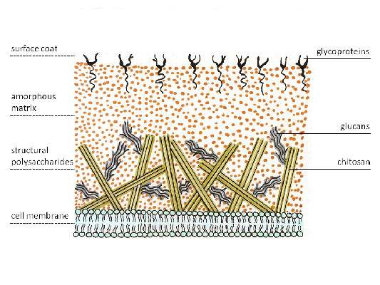
Fig. 4. The cell wall of the fungi.
3. Cell organelles
Actinomycetes, like other bacteria, do not have membrane organelles. Actinomycetes have 70S ribosomes, while fungi have 80S ribosomes, as well as other eukaryotic organisms.
4. Colony growth
The formation of mycelium during growth is what makes actinomycetes more closely related to fungi. In the case of actinomycetes, mycelium is a branching collection of hyphae. Hyphae are divided by partitions into long bacterial cells containing several nucleoids. Partition walls in a number of species can extend in a mutually perpendicular direction. Hyphae branch by budding.
Mycelium growing in a substrate (earth, silt or nutrient medium) is called substrate. It provides the colony with nutrients. Aerial mycelium rises above the substrate, giving the colony “fluffiness” - it forms spores, as well as the so-called “secondary metabolites” (in contrast to the “primary metabolites” of the substrate mycelium), among which there are many antibacterial substances.
Life cycle and physiology of actinomycetes
During the life cycle most of actinomycetes forms spores. Some actinomycetes multiply by fragmentation of the mycelium.
1. Spore formation
Actinomycete spores come from aerial mycelium. These are exospores - they develop outside the mother cell. The hyphae of the aerial mycelium from which spores develop are called sporonos. Disputes can be contained in the thickening at the end of the spore-bearing - sporangia (for example, in streptomycetes, actinoplanes and plimelias), and can be arranged in a chain along the sporangios-carrier (for example, in nocardia and actinomadura).
According to the number of spores formed, actinomycetes are divided into:
- Monospore (for example, Saccaromonospora, Micromonospora, Thermomonospora) - form single spores, more often by budding and subsequent separation by the septum from the mother hypha;
- Oligospore (for example, Actinomadura) - form short chains of spores along the spore bearing;
- Polyspore (most other actinomycetes, for example, Streptomyces, Frankia, Geodermatophilus) - form many spores enclosed in sporangia.
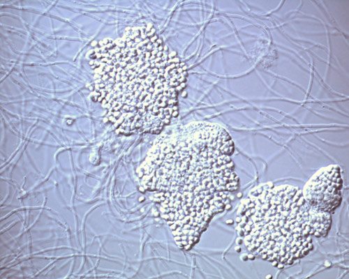
Fig. 5. Sporangium of actinomycetes of the genus Frankia.
Spores of actinomycetes can be mobile - in this case, the spore has a flagellum and can move (spores of actinoplanes, geodermatophiles and dermatophiles). In most cases, spores are immobile and spread by wind, water or animals.

Fig. 6. Dermatophiles, light microscopy.
Sporulation in actinomycetes is particularly active in adverse conditions. The resistance of spores to heating is small compared to the spores of other bacteria, however, they can withstand drying not worse than others, and therefore have tremendous adaptive value. Actinomycetes dominate other microorganisms in dry desert soils.
Germination of a bunch requires a certain humidity of the environment. In the presence of water, the spore swells, enzymes are activated in it and metabolic processes are started, accompanied by the release of growth tubes (future bacterial bodies) and the synthesis of nucleic acids.
2. Type of breath
Most actinomycetes are aerobes (they need oxygen to support life). Optional anaerobes (bacteria that can live both in the presence and in the absence of oxygen) are found among species with a short mycelial stage, propagating by fragmentation of the mycelium.
3. Acid Resistance
Actinomycetes have acid tolerance - stability in an acidic environment, which allows them to live in acid-saturated forest soils. Acid resistance in the laboratory can be determined by the color of the preparation containing actinomycetes according to Zil-Nielsen (fuchsin followed by etching with sulfuric acid and methylene blue). Most actinomycetes with this color do not discolor after acid etching and retain a red fuchsin color. The alkaline environment is unfavorable for these bacteria: at elevated pH they are prone to spore formation.
4. Features of metabolism
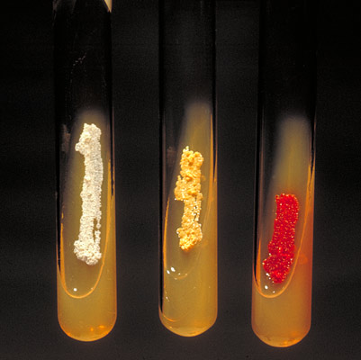
Fig. 7. Aerobic actinomycetes form pigment on mowed agar. From left to right: Actinomadura madurae, Nocardia asteroides, Micromonospora.
The formation of “secondary metabolites” by aerial mycelium was mentioned above. Among them:
- pigments causing various colors of aerial mycelium during growth on media;
- volatile odorous substances that give a characteristic smell to the soil after rain, stagnant water, the skin of some animals;
- antibiotics:
a. antifungal - polyenes;
b. antibacterial - for example, streptomycin, erythromycin, tetracycline, vancomycin;
c. antitumor - anthracyclines, bleomycin.
Where do actinomycetes live?
Actinomycetes are found in the greatest amount in soils, moreover, mycelial forms are much smaller than spores. They play a significant role in the formation of humus, decomposing organic substances that are difficult to utilize by other bacteria. In this regard, actinomycetes are used as sanitary-indicative microorganisms in the sanitary-epidemiological business: the discovery of them in large quantities in soil or water indicates the presence of compost in the corresponding substrate.
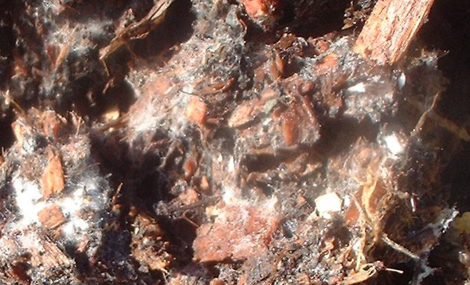
Fig. 8. Actinomycetes in compost.
Actinomycetes are the symbionts of many plants, helping them fix nitrogen. At the same time, many microorganisms of this class are causative agents of plant diseases.
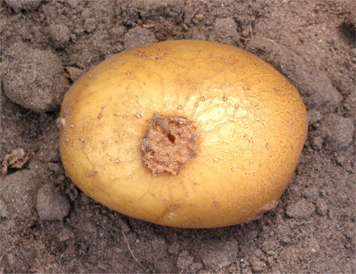
Fig. 9. Streptomycosis of potatoes.
They are also found in the normal microflora of the digestive system of a number of animals, ranging from soil annelids (for example, earthworms) to large livestock.
These microorganisms help break down cellulose, abundantly present in plant foods. In humans, actinomycetes are found in the oral cavity (gums and plaque), intestines (distal parts of the large intestine), on the skin (face, nose wings, behind the ears, between the fingers) and in the organs of the respiratory system (mainly in the upper respiratory tract).

Fig. 10. The microflora of human skin. The type of actinobacteria is indicated by shades of blue, the class of actinomycetes is indicated by bright blue.
Actinomycetes, subject to a decrease in the body's immune reactivity, can cause actinomycosis, an opportunistic disease consisting in the formation of actinomycotic granulomas - clusters of bacterial bodies resembling yellow sulfur grains (“friends”) surrounded by immunocompetent cells. The inflammatory reaction leads to the melting of granulomas, the formation of fistulas, leading to perforations of organs and the spread of bacteria by blood.
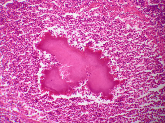
Fig. 11. Actinomycotic druse, Gram stain.
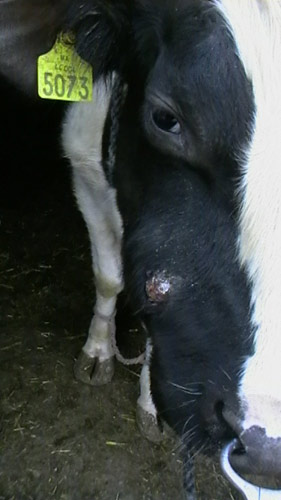
Fig. 12. Actinomycosis of the upper jaw in a cow.
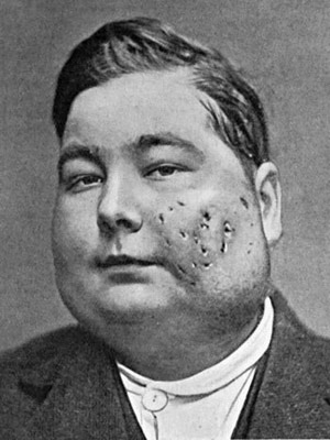
Fig. 13. Maxillary human actinomycosis.
Actinomycetes are amazing organisms that are still misleading many scientists with their similarity to mushrooms. Along with the potential danger in the form of opportunistic actinomycoses, these organisms give people fertile soil and weapons for combating infectious and oncological diseases - antibiotics and cytostatics.